
Catalogue Search | MBRL


Are you sure you want to remove the book from the shelf?

{{itemTitle}}
237
result(s) for
"Albert, Todd J."
Sort by:

What Is the Incidence and Severity of Dysphagia After Anterior Cervical Surgery?
Background
Existing studies suggest a relatively high incidence of dysphagia after anterior cervical decompression and fusion (ACDF). The majority of these studies, however, are retrospective in nature and lack a control group.
Questions/purposes
We therefore (1) prospectively determined the incidence and severity of dysphagia after ACDF using lumbar decompression patients as a control group; and (2) determined which factors, if any, are associated with increased postoperative dysphagia.
Methods
Patients undergoing either one- or two-level ACDF (n = 38) or posterior lumbar decompression (n = 56) were prospectively followed. Baseline patient characteristics were recorded. A dysphagia questionnaire was administered preoperatively and during the 2-week, 6-week, and 12-week postoperative visits. We found no differences in patient age, body mass index, or the preoperative incidence and severity of dysphagia between the cervical and lumbar groups. We compared the incidence and severity of dysphagia between the patients who had cervical and lumbar surgery.
Results
Postoperatively, 71% of patients having cervical spine surgery reported dysphagia at 2 weeks followup. This incidence decreased to 8% at 12 weeks followup. The incidence and severity of dysphagia were greater in the cervical group at 2 and 6 weeks followup with a trend toward greater dysphagia at 12 weeks followup. Body mass index, gender, location of surgery, and the number of surgical levels were not related to the risk of developing dysphagia. We observed a correlation between operative time and the severity of postoperative dysphagia.
Conclusions
Dysphagia is common after ACDF. The incidence and severity of postoperative dysphagia decreases over time, although symptoms may persist at least 12 weeks after surgery.
Level of Evidence
Level II, prospective, comparative study. See Guidelines for Authors for a complete description of levels of evidence.
Journal Article
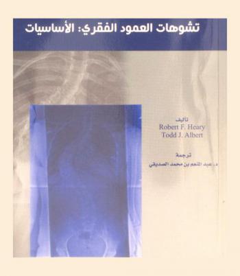
تشوهات العمود الفقرى : الأساسيات
إن هذا الكتاب متميز ومتخصص تخصصا دقيقا في علم انحرافات العمود الفقري وقد حصر جميع المعلومات الدقيقة التي يحتاجها الأطباء المتخصصون بالعظام والعمود الفقري بين دفتيه وهو يمثل عمل مجموعة من أطباء الجراحة العظمية والعصبية يهدف إلى توفير وتحديث الطريقة المثلى لعلاج المرضى الذين يعانون من تشوه في العمود الفقري. اشتمل الكتاب على سبعة وعشرين فصلا يتناول أساسيات جراحة التشوهات البليغة، وقام بتغطية سلسلة من الجراحات الضرورية للتشوه والحالات الأكثر تعقيدا ابتداء من الأطفال إلى الكبار في السن وذلك بالاعتماد على التصوير الشعاعي لتصنيف تشوهات العمود الفقري ومقاطع صور التشوهات السهمية والجبهية كما تم شرح وتوضيح تعقيدات عمليات التشوه الجراحية بعمق ووضوح.
BOOK

Cage height is more important than surface area for preventing subsidence in multilevel anterior cervical discectomy and fusions
Background
The relationships between subsidence and cage dimension and surface area in multilevel anterior cervical discectomy and fusion (ACDF) remain unclear in the current literature. This study aimed to analyze the risk factors influencing cage subsidence following multilevel ACDFs, and the relationship between cage subsidence and cage dimensions, surface area, patient-reported outcomes, and fusion rates.
Methods
Patients who had undergone primary multilevel ACDFs with a minimum follow-up duration of 1 year were enrolled in this study. Cage subsidence was evaluated using lateral radiographs and defined as a decrease in disc height ≥3 mm. The patients were classified into the cage subsidence and no cage subsidence groups. Neck Disability Index (NDI) scores were recorded at baseline and every clinic visit. Fusion rates were evaluated at the final follow-up.
Results
Eighty-five patients (mean age, 58.6 years; mean follow-up, 12.7 months) were included. The cage subsidence rates were 5%, 12%, 23%, and 31% at the 6-week, 3-month, 6-month, and 1-year follow-up, respectively. The demographic, perioperative, and radiographic parameters were not different between the two groups. Cage type, width, depth, and surface area did not influence the subsidence rates. Cage height was positively correlated with the depth of cage subsidence (
p
< 0.001,
R
= 0.263). A cage height ≥8 mm was associated with a higher risk of cage subsidence (odds ratio: 3.9, 95% confidence interval: 1.8–8.5,
p
< 0.001). An SPA ≥ 4° of the distal screw was identified as a risk factor for cage subsidence at the distal level. The overall fusion rate was 88.5% in both groups. NDI scores did not differ preoperatively or at follow-up.
Conclusions
Cage height and cage subsidence were significantly correlated in ACDF patients. A cage height ≥8 mm may be a risk factor for cage subsidence. A cage with a larger surface area may not provide protection against subsidence.
Journal Article

Memorabilia ; Oeconomicus
\"Xenophon (ca. 430 to ca. 354 BCE), a member of a wealthy but politically quietist Athenian family and an admirer of Socrates, left Athens in 401 BCE to serve as a mercenary commander for Cyrus the Younger of Persia, then joined the staff of King Agesilaus II of Sparta before settling in Elis and, in the aftermath of the battle of Leuctra in 371 BCE, retiring to Corinth. His historical and biographical works, Socratic dialogues and reminiscences, and short treatises on hunting, horsemanship, economics, and the Spartan constitution are richly informative about his own life and times. This volume collects Xenophon's portrayals of his associate, Socrates. In Memorabilia (or Memoirs of Socrates) and in Oeconomicus, a dialogue about household management, we see the philosopher through Xenophon's eyes. Here, as in the accompanying Symposium, we also obtain insight on life in Athens. The volume concludes with Xenophon's Apology, an interesting complement to Plato's account of Socrates's defense at his trial.\" -- Publisher website.
BOOK

The Spine Physical Examination Using Telemedicine: Strategies and Best Practices
Study Design:
Technical note.
Objectives:
To provide spine surgeons new to telemedicine with a structured physical examination technique based on manual motor testing principles.
Methods:
Expert experience describing a series of specific maneuvers for upper and lower extremity strength testing that can be performed using a telemedicine platform. In addition, we offer instruction on “setting up” for these visits and highlight special tests that can be used to diagnose specific cervical and lumbar spine conditions.
Results:
From our experiences in conducting telemedicine visits, we provide a means of testing and scoring upper and lower extremity strength for interpretation of weakness in the context of traditional manual motor testing. Also, we acknowledge the limitations of a remote examination and discuss maneuvers that cannot be performed remotely.
Conclusions:
COVID-19 has drastically altered the delivery of care for patients with spine-related complaints. The need for social distancing has led to the widespread adoption of telemedicine. This technical note provides an urgently needed framework for the standardization of the remote physical exam. Validation of the exam as a diagnostic tool will be a crucial next step in studying the impact of telemedicine.
Journal Article

Management of Odontoid Fractures in the Elderly: A Review of the Literature and an Evidence-Based Treatment Algorithm
Abstract
Odontoid fractures are the most common fracture of the axis and the most common cervical spine fracture in patients over 65. Despite their frequency, there is considerable ambiguity regarding optimal management strategies for these fractures in the elderly. Poor bone health and medical comorbidities contribute to increased surgical risk in this population; however, nonoperative management is associated with a risk of nonunion or fibrous union. We provide a review of the existing literature and discuss the classification and evaluation of odontoid fractures. The merits of operative vs nonoperative management, fibrous union, and the choice of operative approach in elderly patients are discussed. A treatment algorithm is presented based on the available literature. We believe that type I and type III odontoid fractures can be managed in a collar in most cases. Type II fractures with any additonal risk factors for nonunion (displacement, comminution, etc) should be considered for surgical management. However, the risks of surgery in an elderly population must be carefully considered on a case-by-case basis. In a frail elderly patient, a fibrous nonunion with close follow-up is an acceptable outcome. If operative management is chosen, a posterior approach is should be chosen when fracture- or patient-related factors make an anterior approach challenging. The high levels of morbidity and mortality associated with odontoid fractures should encourage all providers to pursue medical co-management and optimization of bone health following diagnosis.
Journal Article

Comparison of Transcranial Electric Motor and Somatosensory Evoked Potential Monitoring During Cervical Spine Surgery
BACKGROUND:There has been little enthusiasm for somatosensory evoked potential monitoring in cervical spine surgery as a result, in part, of the increased risk of motor tract injury at this level, to which somatosensory monitoring may be insensitive. Transcranial electric motor evoked potential monitoring allows assessment of the motor tracts; therefore, we compared transcranial electric motor evoked potential and somatosensory evoked potential monitoring during cervical spine surgery to determine the temporal relationship between the changes in the potentials demonstrated by each type of monitoring and neurological sequelae and to identify patient-related and surgical factors associated with intraoperative neurophysiological changes.
METHODS:Somatosensory evoked potential and transcranial electric motor evoked potential data recorded for 427 patients undergoing anterior or posterior cervical spine surgery between January 1999 and March 2001 were analyzed. All patients who showed substantial (at least 60%) or complete unilateral or bilateral amplitude loss, for at least ten minutes, during the transcranial electric motor evoked potential and/or somatosensory evoked potential monitoring were identified.
RESULTS:Twelve of the 427 patients demonstrated substantial or complete loss of amplitude of the transcranial electric motor evoked potentials. Ten of those patients had complete reversal of the loss following prompt intraoperative intervention, whereas two awoke with a new motor deficit. Somatosensory evoked potential monitoring failed to identify any change in one of the two patients, and the change in the somatosensory evoked potentials lagged behind the change in the transcranial electric motor evoked potentials by thirty-three minutes in the other. No patient showed loss of amplitude of the somatosensory evoked potentials in the absence of changes in the transcranial electric motor evoked potentials. Transcranial electric motor evoked potential monitoring was 100% sensitive and 100% specific, whereas somatosensory evoked potential monitoring was only 25% sensitive; it was, however, 100% specific.
CONCLUSIONS:Transcranial electric motor evoked potential monitoring appears to be superior to conventional somatosensory evoked potential monitoring for identifying evolving motor tract injury during cervical spine surgery. Surgeons should strongly consider using this modality when operating on patients with cervical spondylotic myelopathy in general and on those with ossification of the posterior longitudinal ligament in particular.
LEVEL OF EVIDENCE:Diagnostic study, Level I-1 (testing of previously developed diagnostic criteria in series of consecutive patients [with universally applied reference “gold” standard]). See Instructions to Authors for a complete description of levels of evidence.
Journal Article

Chondrocyte‐like nested cells in the aged intervertebral disc are late‐stage nucleus pulposus cells
Aging is a major risk factor of intervertebral disc degeneration and a leading cause of back pain. Pathological changes associated with disc degeneration include the absence of large, vacuolated and reticular‐shaped nucleus pulposus cells, and appearance of smaller cells nested in lacunae. These small nested cells are conventionally described as chondrocyte‐like cells; however, their origin in the intervertebral disc is unknown. Here, using a genetic mouse model and a fate mapping strategy, we have found that the chondrocyte‐like cells in degenerating intervertebral discs are, in fact, nucleus pulposus cells. With aging, the nucleus pulposus cells fuse their cell membranes to form the nested lacunae. Next, we characterized the expression of sonic hedgehog (SHH), crucial for the maintenance of nucleus pulposus cells, and found that as intervertebral discs age and degenerate, expression of SHH and its target Brachyury is gradually lost. The results indicate that the chondrocyte‐like phenotype represents a terminal stage of differentiation preceding loss of nucleus pulposus cells and disc collapse. Lineage tracing of neonatal mouse nucleus pulposus (NP) cells of the intervertebral disc using Krt19CreERT; R26mT/mG line shows that reticular‐shaped NP cells differentiate into the chondrocyte‐like cell (CLCs) with aging. The study also shows that the nested CLCs are formed by the fusion of several NP cells. And differentiation of NP into CLC was associated with a decline in the expression of SHH and Brachyury.
Journal Article

Deviation analysis of C2 translaminar screw placement assisted by a novel rapid prototyping drill template: a cadaveric study
Purpose
The goal of this study is to evaluate the accuracy of patient-specific CT-based rapid prototype drill templates for C2 translaminar screw insertion.
Methods
Volumetric CT scanning was performed in 32 cadaveric cervical spines. Using computer software, the authors constructed drill templates that fit onto the posterior surface of the C2 vertebrae with drill guides to match the slope of the patient’s lamina. Thirty-two physical templates were created from the computer models using a rapid prototyping machine. The drill templates were used to guide drilling of the lamina and post-operative CT images were obtained. The entry point and direction of the planned and inserted screws were measured and compared.
Results
Sixty-four C2 translaminar screws were placed without violating the cortical bone of a single lamina. The bilateral average transverse angle of intended and actual screw for C2TLS was 56.60 ± 2.22°, 56.38 ± 2.51°, 56.65 ± 2.24°, 56.39 ± 2.45°. The bilateral mean coronal angle of the planned and actual screw for C2TLS was 0°, 0°, −0.07 ± 0.32°, 0.12 ± 0.57°. The average displacement of the entry point of the superior and inferior C2TLS in the
x
,
y
,
z
axis was 0.27 ± 0.85, 0.49 ± 1.46, −0.28 ± 0.69, 0.43 ± 0.88, 0.38 ± 1.51, 0.23 ± 0.64 mm.
Conclusion
The small deviations seen are likely due to human error in the form of small variations in the surgical technique and use of software to design the prototype. This technology improves the safety profile of this fixation technique and should be further studied in clinical applications.
Journal Article