Catalogue Search | MBRL


Are you sure you want to remove the book from the shelf?

{{itemTitle}}
44
result(s) for
"Grimm, Jochen"
Sort by:
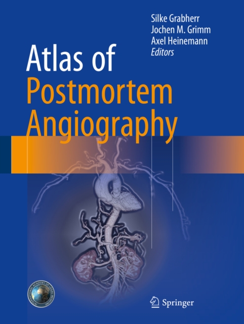
Atlas of postmortem angiography
This atlas of postmortem angiography provides a summary of techniques that have been developed and used in order to visualize the human vascular system.The indications, advantages, limitations, and pitfalls of the different techniques are explained in detail through the use of examples from real cases and a wealth of informative images, as well.
eBook

Age determination of vessel wall hematoma in spontaneous cervical artery dissection: A multi-sequence 3T Cardiovascular Magnetic resonance study
Background
Previously proposed classifications for carotid plaque and cerebral parenchymal hemorrhages are used to estimate the age of hematoma according to its signal intensities on T1w and T2w MR images. Using these classifications, we systematically investigated the value of cardiovascular magnetic resonance (CMR) in determining the age of vessel wall hematoma (VWH) in patients with spontaneous cervical artery dissection (sCAD).
Methods
35 consecutive patients (mean age 43.6 ± 9.8 years) with sCAD received a cervical multi-sequence 3T CMR with fat-saturated black-blood T1w-, T2w- and TOF images. Age of sCAD was defined as time between onset of symptoms (stroke, TIA or Horner's syndrome) and the CMR scan. VWH were categorized into hyperacute, acute, early subacute, late subacute and chronic based on their signal intensities on T1w- and T2w images.
Results
The mean age of sCAD was 2.0, 5.8, 15.7 and 58.7 days in patients with acute, early subacute, late subacute and chronic VWH as classified by CMR (p < 0.001 for trend). Agreement was moderate between VWH types in our study and the previously proposed time scheme of signal evolution for cerebral hemorrhage, Cohen's kappa 0.43 (p < 0.001). There was a strong agreement of CMR VWH classification compared to the time scheme which was proposed for carotid intraplaque hematomas with Cohen's kappa of 0.74 (p < 0.001).
Conclusions
Signal intensities of VWH in sCAD vary over time and multi-sequence CMR can help to determine the age of an arterial dissection. Furthermore, findings of this study suggest that the time course of carotid hematomas differs from that of cerebral hematomas.
Journal Article

Performance of post-mortem CT compared to autopsy in children
Introduction
Radiological techniques such as non-enhanced post-mortem computed tomography (PMCT) play an increasingly important role in death investigations, especially in cases of non-medicolegal context of death, where the consent of the next of kin is required to perform autopsy. Such consent is often difficult to obtain for deceased children, and radiological methods may be an acceptable alternative. The aim of our study was to evaluate the performance of PMCT explorations compared to medicolegal conventional autopsies in children and its potential usefulness in non-medicolegal situations.
Methods
We retrospectively reviewed a group of 26 children aged 0–12 years who died of different causes, which were investigated by both conventional autopsy and PMCT. We compared the findings extracted from radiological and autopsy reports. All findings were grouped according to their importance with respect to cause of death and to the anatomical structure they covered: organs, vascular system, soft tissue, and skeletal system.
Results
A significantly larger number of findings were detected by autopsy compared to PMCT. Autopsy proved to be superior to PMCT, notably at detecting organ, soft tissue, and vascular findings, while PMCT was superior at detecting bone findings. However, no statistically significant differences were found between the methods concerning the essential findings used to define the cause of death.
Conclusions
In children, PMCT was less sensitive than conventional autopsy for detecting general findings. However, most essential findings were detected by both methods. PMCT was superior to autopsy for the detection of bone lesions in children.
Advances in knowledge
Up to today, very rare literature exists concerning PMCT in children, especially in a forensic setting. This article investigates the advantages and limitations of PMCT compared to autopsy in a unique study group and discusses possibilities for future developments.
Journal Article

Tetravalent Influenza Vaccine Is Not Associated With Neuroaxonal Damage in Multiple Sclerosis Patients
BackgroundEfficacy of vaccines and disease activity linked to immunization are major concerns among people with multiple sclerosis (pwMS).ObjectiveTo assess antibody responses to seasonal influenza antigens and vaccine-associated neuroaxonal damage utilizing serum neurofilament light chain (sNfL) in pwMS receiving dimethyl fumarate (DMF).MethodsIn this prospective study, the 2020/2021 seasonal tetravalent influenza vaccine was administered to 20 pwMS treated with DMF and 15 healthy controls (HCs). The primary endpoints were responder rate of strain-specific antibody production (seroconversion or significant (4-fold) increase in influenza-antibody titers for ≥2/4 strains) at 30 days post-vaccination and changes in sNfL levels.ResultsAll patients treated with DMF fulfilled the responder criteria for immunization compared with 53% of the controls. However, higher proportions of HCs already had influenza-antibody titers ≥1:40 at baseline (53% vs. 41%, p = 0.174). sNfL levels were comparable among both groups at baseline and did not increase 34 days after vaccination. In addition, no clinical or radiological disease reactivation was found.ConclusionDMF-treated patients mount an adequate humoral immune response to influenza vaccines. Within the limits of the small cohort investigated, our data suggest that influenza immunization is not associated with clinical or subclinical disease reactivation.
Journal Article
Pathomorphological and CT-angiographical characteristics of coronary atherosclerotic plaques in cases of sudden cardiac death
The goal of this study was to assess the localization and types of thrombosed plaques in cases of sudden cardiac death attributed to coronary artery disease and to evaluate possible correlations with body mass index (BMI) and increased heart weight. This retrospective study was performed on forensic cases for which the cause of death was attributed to coronary artery disease. A complete autopsy and a multi-phase postmortem computed tomography (CT) angiography (MPMCTA) were performed in all cases. Eighty-five cases were selected (mean age, 55.18 ± 11.04 years; 72 men and 13 women). MPMCTA performed prior to autopsy enabled an evaluation of coronary artery perfusion before dissection of the body and helped therefore to guide sampling for histology. An acute coronary thrombosis was found in 57 cases, which included plaque erosion in 26 cases (mean age, 46.73 ± 8.33 years) and rupture or intra-plaque hemorrhage in 31 cases (mean age, 58.23 ± 10.62 years). Erosions were most frequently found in the left anterior descending artery (61.5 %), while only 35.48 % of ruptures were observed in this artery. Chronic coronary pathology was considered as the main cause of death in 28 cases (mean age, 59.64 ± 9.47 years). Sixty-two of the cases (72.94 %) had a BMI in the overweight category (BMI ≥25), with the highest mean BMI in patients with chronic coronary pathology without acute thrombosis found at autopsy. The heart weight was above the predicted reference values in 52 cases (61.18 %). Our results are in accordance with previously published studies on the spatial distribution of vulnerable plaques. We observed a higher percentage of eroded plaques than previously reported. Patients with coronary erosions were significantly younger than those with plaque rupture or those without an acute coronary thrombosis (
p
values <0.0001). BMI and heart weight were significantly higher for cases without thrombosis in comparison with those with plaque rupture (
p
values 0.028 and 0.003, respectively). Our results indicating that increased BMI and overweight hearts are associated with chronic ischemic heart disease are compatible with clinical studies. Performing more postmortem studies on forensic autopsies, including modern radiological examinations with MPMCTA, can enhance the detection of vulnerable plaques in living patients and prevent sudden cardiac death.
Journal Article

Pituitary Apoplexy in Macroadenoma After Minor Surgery: An Unusual Case and Literature Review
Pituitary apoplexy is a rare and severe complication of pituitary adenoma that may present with new-onset headache, ocular palsy, visual disturbances, life-threatening electrolyte imbalance, and endocrinological disturbances due to pituitary hemorrhage and/or infarction. We report the case of a 58-year-old previously healthy patient who developed isolated mild oculomotor nerve palsy of the left eye following osteosynthesis of a traumatic right distal radius fracture. Initial cerebral magnetic resonance imaging showed a pituitary macroadenoma without characteristic signs of pituitary infarction or hemorrhage. The patient presented to the neurology department on the fifth postoperative day with malaise and fatigue due to pituitary insufficiency, deteriorated rapidly and required intensive care monitoring. Clinical stabilization was achieved through the administration of hydrocortisone, and transsphenoidal resection of the pituitary lesion was performed on the 10th day after acute symptom onset. Histological examination revealed a necrotic pituitary adenoma. Pituitary apoplexy may occur after minor surgery in patients with pituitary adenoma. Clinicians should pay particular attention to laboratory signs of pituitary insufficiency in new-onset oculomotor nerve palsy associated with sellar lesions, as cerebral imaging may miss pituitary apoplexy and therefore delay diagnosis and treatment. In our case, delayed decompressive transsphenoidal resection resulted in the normalization of the oculomotor nerve palsy while the pituitary insufficiency persisted. The potential impact of an earlier surgical intervention on the outcome of pituitary function remains uncertain.
Journal Article

Neck-MRI experience for investigation of survived strangulation victims
For the medicolegal evaluation of victims of survived strangulation, a neck-magnetic resonance imaging (MRI) can be performed for assessing lesions in the inner soft tissues (fat, muscles or lymph nodes, for example). In our institute, such MRI examinations have been performed for a test period of 4 years with the aim of evaluating the use of this tool by forensic pathologists and identifying medicolegal indicators for the performance of neck-MRI in surviving victims of strangulation. We retrospectively reviewed medicolegal reports from all victims examined during the test period. We extracted objective lesions (e.g. petechiae, bruising and abrasions) and reported clinical symptoms (e.g. vision disorder, dysphasia) from the reports. These findings were compared to those reported from the neck-MRI. In total, 112 victims were clinically examined after suspected strangulation. Eleven of these victims underwent an MRI examination of the neck. Eighty-four of the victims presented objective lesions during the clinical examination, with eight showing signs of both petechiae and bruising. Neck-MRI was performed in four of these eight victims and three of them showed lesions visible in MRI. Of 76 victims with bruising as the only objective finding, 66 victims described clinical symptoms. Of those 66 victims, seven were examined by MRI and two demonstrated lesions in MRI. When MRI was performed, relevant findings were detected in 45% of the cases. This leads to the suspicion that many more findings could have been detected in the other victims, if an MRI had been performed in those cases. Our results lead us to the conclusion that an MRI examination of victims of suspected strangulation is useful, and strict indications for its application should be established.
Journal Article

Forensic radiology : Introduction and overview
In forensic medicine, documentation of findings is essential. During an autopsy, this is usually achieved by photography. However, there are numerous injuries that remain undetected even during a classic autopsy. In recent years, the importance of forensic radiology has grown in many countries to improve the documentation of findings and to increase the quality of post-mortem examinations.
While many methods, such as conventional X‑rays or computed tomography, can be transferred quite easily to the post-mortem field, there are other methods that are more difficult to adapt. For example, performing a post-mortem angiography requires a specific concept that allows the vascular system to be filled and a contrast agent to circulate. Performing post-mortem magnetic resonance imaging is also a challenge, as image contrast depends on the temperature of the body being examined. When applying forensic radiology on living persons in the field of \"clinical forensic medicine\", there are further elements to consider. In particular, the question arises if radiological methods are acceptable for purely forensic medical purposes without a clinical indication.
This overview article is intended to explain the various methods of forensic radiology, their areas of application, and their advantages and disadvantages. It also describes important historical developments in the use of forensic radiology and its current spread in German-speaking countries as well as current and future developments. Thanks to this information and a summarizing overview table, clear indications and recommendations for the use of forensic radiology in practice can be obtained.
Journal Article
Computed tomography angiography vs 3 T black-blood cardiovascular magnetic resonance for identification of symptomatic carotid plaques
Background
The purpose of this prospective study was to perform a head-to-head comparison of the two methods most frequently used for evaluation of carotid plaque characteristics: Multi-detector Computed Tomography Angiography (MDCTA) and black-blood 3 T-cardiovascular magnetic resonance (bb-CMR) with respect to their ability to identify symptomatic carotid plaques.
Methods
22 stroke unit patients with unilateral symptomatic carotid disease and >50% stenosis by duplex ultrasound underwent MDCTA and bb-CMR (TOF, pre- and post-contrast fsT1w-, and fsT2w- sequences) within 15 days of symptom onset. Both symptomatic and contralateral asymptomatic sides were evaluated. By bb-CMR, plaque morphology, composition and prevalence of complicated AHA type VI lesions (AHA-LT6) were evaluated. By MDCTA, plaque type (non-calcified, mixed, calcified), plaque density in HU and presence of ulceration and/or thrombus were evaluated. Sensitivity (SE), specificity (SP), positive and negative predictive value (PPV, NPV) were calculated using a 2-by-2-table.
Results
To distinguish between symptomatic and asymptomatic plaques AHA-LT6 was the best CMR variable and presence / absence of plaque ulceration was the best CT variable, resulting in a SE, SP, PPV and NPV of 80%, 80%, 80% and 80% for AHA-LT6 as assessed by bb-CMR and 40%, 95%, 89% and 61% for plaque ulceration as assessed by MDCTA. The combined SE, SP, PPV and NPV of bb-CMR and MDCTA was 85%, 75%, 77% and 83%, respectively.
Conclusions
Bb-CMR is superior to MDCTA at identifying symptomatic carotid plaques, while MDCTA offers high specificity at the cost of low sensitivity. Results were only slightly improved over bb-CMR alone when combining both techniques.
Journal Article
Comparison of symptomatic and asymptomatic atherosclerotic carotid plaques using parallel imaging and 3 T black-blood in vivo CMR
To determine if black-blood 3 T cardiovascular magnetic resonance (bb-CMR) can depict differences between symptomatic and asymptomatic carotid atherosclerotic plaques in acute ischemic stroke patients.
In this prospective monocentric observational study 34 patients (24 males; 70 ±9.3 years) with symptomatic carotid disease defined as ischemic brain lesions in one internal carotid artery territory on diffusion weighted images underwent a carotid bb-CMR at 3 T with fat-saturated pre- and post-contrast T1w-, PDw-, T2w- and TOF images using surface coils and Parallel Imaging techniques (PAT factor = 2) within 10 days after symptom onset. All patients underwent extensive clinical workup (lab, brain MR, duplex sonography, 24-hour ECG, transesophageal echocardiography) to exclude other causes of ischemic stroke. Prevalence of American Heart Association lesion type VI (AHA-LT6), status of the fibrous cap, presence of hemorrhage/thrombus and area measurements of calcification, necrotic core and hemorrhage were determined in both carotid arteries in consensus by two reviewers who were blinded to clinical information. McNemar and Wilcoxon's signed rank tests were use for statistical comparison. A p-value <0.05 was considered statistically significant.
Symptomatic plaques showed a higher prevalence of AHA-LT6 (67.7% vs. 11.8%; p < 0.001; odds ratio = 12.5), ruptured fibrous caps (44.1% vs. 2.9%; p < 0.001; odds ratio = 15.0), juxtaluminal thrombus (26.5 vs. 0%; p < 0.01; odds ratio = 7.3) and intraplaque hemorrhage (58.6% vs. 11.8%; p = 0.01; odds ratio = 3.8). Necrotic core and hemorrhage areas were greater in symptomatic plaques (14.1 mm2 vs. 5.5 mm2 and 13.6 mm2 vs. 5.3 mm2; p < 0.01, respectively).
3 T bb-CMR is able to differentiate between symptomatic and asymptomatic carotid plaques, demonstrating the potential of bb-CMR to differentiate between stable and vulnerable lesions and ultimately to identify patients with low versus high risk for cardiovascular complications. Best predictors of the symptomatic side were a ruptured fibrous cap, AHA-LT 6, juxtaluminal hemorrhage/thrombus, and intraplaque hemorrhage.
Journal Article