Catalogue Search | MBRL


Are you sure you want to remove the book from the shelf?

{{itemTitle}}
45
result(s) for
"Kretsinger, Robert H"
Sort by:


Molecular Dynamics Study of the Changes in Conformation of Calmodulin with Calcium Binding and/or Target Recognition
Calmodulin is a calcium binding protein with two lobes, N-lobe and C-lobe, which evolved from duplication and fusion of a single precursor lobe of a pair of EF-hand. These two lobes of calmodulin show subtle differences in calcium binding and target recognition; these are important for the functions of calmodulin. Since the structures, especially main chain conformations, of two EF-lobes in holo-form are quite similar; this is a good example to evaluate the effect of side chains for structural dynamics. We analyzed the structure of calmodulin using molecular dynamics and found differences in conformational ensembles between N- and C-lobes. We also showed the mutant structures created by homology modeling could reproduce the difference of dynamic motion between N- and C-lobes.
Journal Article

Shrinking of repeating unit length in leucine-rich repeats from double-stranded DNA viruses
Leucine-rich repeats (LRRs) are present in over 563,000 proteins from viruses to eukaryotes. LRRs repeat in tandem and have been classified into fifteen classes in which the repeat unit lengths range from 20 to 29 residues. Most LRR proteins are involved in protein-protein or ligand interactions. The amount of genome sequence data from viruses is increasing rapidly, and although viral LRR proteins have been identified, a comprehensive sequence analysis has not yet been done, and their structures, functions, and evolution are still unknown. In the present study, we characterized viral LRRs by sequence analysis and identified over 600 LRR proteins from 89 virus species. Most of these proteins were from double-stranded DNA (dsDNA) viruses, including nucleocytoplasmic large dsDNA viruses (NCLDVs). We found that the repeating unit lengths of 11 types are one to five residues shorter than those of the seven known corresponding LRR classes. The repeating units of six types are 19 residues long and are thus the shortest among all LRRs. In addition, two of the LRR types are unique and have not been observed in bacteria, archae or eukaryotes. Conserved strongly hydrophobic residues such as Leu, Val or Ile in the consensus sequences are replaced by Cys with high frequency. Phylogenetic analysis indicated that horizontal gene transfer of some viral LRR genes had occurred between the virus and its host. We suggest that the shortening might contribute to the survival strategy of viruses. The present findings provide a new perspective on the origin and evolution of LRRs.
Journal Article

Super Secondary Structure Consisting of a Polyproline II Helix and a β-Turn in Leucine Rich Repeats in Bacterial Type III Secretion System Effectors
Leucine rich repeats (LRRs) are present in over 100,000 proteins from viruses to eukaryotes. The LRRs are 20–30 residues long and occur in tandem. LRRs form parallel stacks of short β-strands and then assume a super helical arrangement called a solenoid structure. Individual LRRs are separated into highly conserved segment (HCS) with the consensus of LxxLxLxxNxL and variable segment (VS). Eight classes have been recognized. Bacterial LRRs are short and characterized by two prolines in the VS; the consensus is xxLPxLPxx with Nine residues (N-subtype) and xxLPxxLPxx with Ten residues (T-subtype). Bacterial LRRs are contained in type III secretion system effectors such as YopM, IpaH3/9.8, SspH1/2, and SlrP from bacteria. Some LRRs in decorin, fribromodulin, TLR8/9, and FLRT2/3 from vertebrate also contain the motifs. In order to understand structural features of bacterial LRRs, we performed both secondary structures assignments using four programs—DSSP-PPII, PROSS, SEGNO, and XTLSSTR—and HELFIT analyses (calculating helix axis, pitch, radius, residues per turn, and handedness), based on the atomic coordinates of their crystal structures. The N-subtype VS adopts a left handed polyproline II helix (PPII) with four, five or six residues and a type I β-turn at the C-terminal side. Thus, the N-subtype is characterized by a super secondary structure consisting of a PPII and a β-turn. In contrast, the T-subtype VS prefers two separate PPIIs with two or three and two residues. The HELFIT analysis indicates that the type I β-turn is a right handed helix. The HELFIT analysis determines three unit vectors of the helix axes of PPII (P), β-turn (B), and LRR domain (A). Three structural parameters using these three helix axes are suggested to characterize the super secondary structure and the LRR domain.
Journal Article
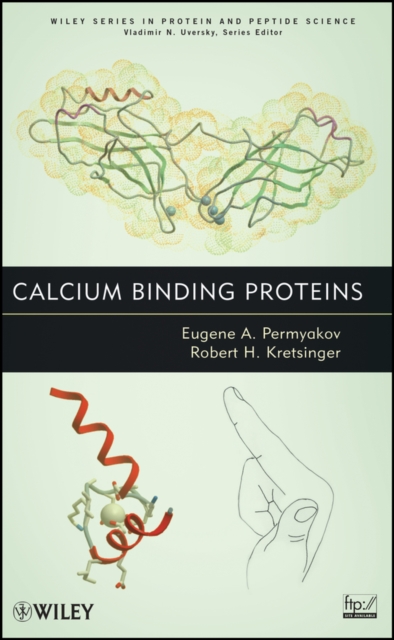
Calcium binding proteins
Calcium Binding Proteins explains the unique and highly diverse functions of calcium in biology, which are realized by calcium binding proteins. The structures and physical characteristics of these calcium binding proteins are described, as well as their functions and general patterns of their evolution. Techniques that underlie the description of proteins are discussed, including NMR, circular dichroism, optical rotatory dispersion spectroscopy, calorimetry,and crystallography. The book discusses the patterns of bochmical phenomena such as calcium homeostasis, mineralization, and cell signaling that involve specific proteins. It summarizes ongoing research and presents general hypotheses that help to focus future research, and also provides a conceptual framework and a description of the underlying techniques that permits someone entering the field to become conversant.
eBook


Two-Dimensional Crystal Structures of Protein Kinase C- δ, Its Regulatory Domain, and the Enzyme Complexed with Myelin Basic Protein
Two-dimensional crystals of protein kinase C (PKC)
δ, its regulatory domain (RD
δ), and the enzyme complexed with the substrate myelin basic protein have been grown on lipid monolayers composed of phosphatidylcholine: phosphatidylserine: diolein (45:50:5, molar ratio). Images have been reconstructed to 10-Å resolution. The unit cells of all three proteins have cell edges a
=
b and interedge angle
γ
=
60
o. RD
δ has an edge length of 33
±
1
Å, and its reconstruction is donut shaped. The three-dimensional reconstructions from the PKC
δ C1b crystal structure (Zhang et al., 1995) can be accommodated in this two-dimensional projection. Intact PKC
δ has an edge length of 46
±
1
Å in the presence or absence of a nonhydrolyzable ATP analog, AMP-PnP. Its reconstruction has a similar donut shape, which can accommodate the C1b domain, but the spacing between donuts is greater than that in RD
δ; some additional structure is visible between the donuts. The complex of PKC
δ and myelin basic protein, with or without AMP-PnP, has an edge length of 43
±
1
Å and a distinct structure. These results indicate that the C1 domains of RD
δ are tightly packed in the plane of the membrane in the two-dimensional crystals, that there is a single molecule of PKC
δ in the unit cell, and that its interaction with myelin basic protein induces a shift in conformation and/or packing of the enzyme.
Journal Article


Comparative Geometrical Analysis of Leucine-Rich Repeat Structures in the Nod-Like and Toll-Like Receptors in Vertebrate Innate Immunity
The NOD-like receptors (NLRs) and Toll-like receptors (TLRs) are pattern recognition receptors that are involved in the innate, pathogen pattern recognition system. The TLR and NLR receptors contain leucine-rich repeats (LRRs) that are responsible for ligand interactions. In LRRs short β-strands stack parallel and then the LRRs form a super helical arrangement of repeating structural units (called a coil of solenoids). The structures of the LRR domains of NLRC4, NLRP1, and NLRX1 in NLRs and of TLR1-5, TLR6, TLR8, TLR9 in TLRs have been determined. Here we report nine geometrical parameters that characterize the LRR domains; these include four helical parameters from HELFIT analysis. These nine parameters characterize well the LRR structures in NLRs and TLRs; the LRRs of NLR adopts a right-handed helix. In contrast, the TLR LRRs adopt either a left-handed helix or are nearly flat; RP105 and CD14 also adopt a left-handed helix. This geometrical analysis subdivides TLRs into four groups consisting of TLR3/TLR8/TLR9, TLR1/TLR2/TRR6, TLR4, and TLR5; these correspond to the phylogenetic tree based on amino acid sequences. In the TLRs an ascending lateral surface that consists of loops connecting the β-strand at the C-terminal side is involved in protein, protein/ligand interactions, but not the descending lateral surface on the opposite side.
Journal Article
Troponin and Parvalbumin Calcium Binding Regions Predicted in Myosin Light Chain and T4 Lysozyme
A computer search of available protein sequences and structures suggests that bacteriophage T4 lysozyme contains one region and that rabbit myosin light chains contain three regions similar, and supposedly homologous, to the calcium binding region of carp muscle calcium binding parvalbumin.
Journal Article