Catalogue Search | MBRL


Are you sure you want to remove the book from the shelf?

{{itemTitle}}
98
result(s) for
"Samei, Ehsan"
Sort by:

Characterizing imaging radiation risk in a population of 8918 patients with recurrent imaging for a better effective dose
An updated extension of effective dose was recently introduced, namely relative effective dose (
E
r
), incorporating age and sex factors. In this study we extended
E
r
application to a population of about 9000 patients who underwent multiple CT imaging exams, and we compared it with other commonly used radiation protection metrics in terms of their correlation with radiation risk. Using Monte Carlo methods,
E
r
, dose-length-product based effective dose (
E
DLP
), organ-dose based effective dose (
E
OD
), and organ-dose based risk index (
RI
) were calculated for each patient. Each metric’s dependency to
RI
was assessed in terms of its sensitivity and specificity.
E
r
showed the best sensitivity, specificity, and agreement with
RI
(R
2
= 0.97); while
E
DLP
yielded the lowest specificity and, along with
E
OD
, the lowest sensitivity. Compared to other metrics,
E
r
provided a closer representation of patient and group risk also incorporating age and sex factors within the established framework of effective dose.
Journal Article
An in silico evaluation of signal and separability properties of k-edge materials in spectral CT
Spectral CT can acquire signal at multiple x-ray energy levels. This enables material quantification by exploiting differences in x-ray attenuation across energy levels, particularly for k-edge materials. This simulation study quantified the signal and separability of current and potential clinical contrast agents across a range of materials and energies. A validated CT simulation platform was used to simulate a clinical photon-counting CT scanner with two energy thresholds. A cylindrical phantom containing common biological materials, clinical contrast agents, candidate contrast agents and nanoparticles, and investigational materials was imaged with varying upper energy thresholds (50–90 keV). At each energy level, images were assessed for noise, each material was assessed for contrast, and each material pair was evaluated for separability. Material contrasts reached peak value at the closest threshold higher than their respective k-edge. The energy threshold that produced the highest separability for each pair was characterized. Selection of energy threshold was dependent on the materials of interest. Threshold values at or just above a material’s k-edge maximized material signal while separability was maximized by the threshold that best separated k-edge signals.
Journal Article

Comparison of 12 surrogates to characterize CT radiation risk across a clinical population
Objectives
Quantifying radiation burden is essential for justification, optimization, and personalization of CT procedures and can be characterized by a variety of risk surrogates inducing different radiological risk reflections. This study compared how twelve such metrics can characterize risk across patient populations.
Methods
This study included 1394 CT examinations (abdominopelvic and chest). Organ doses were calculated using Monte Carlo methods. The following risk surrogates were considered: volume computed tomography dose index (CTDI
vol
), dose-length product (DLP), size-specific dose estimate (SSDE), DLP-based effective dose (ED
k
), dose to a defining organ (OD
D
), effective dose and risk index based on organ doses (ED
OD
, RI), and risk index for a 20-year-old patient (RI
rp
). The last three metrics were also calculated for a reference ICRP-110 model (OD
D,0
, ED
0
, and RI
0
). Lastly, motivated by the ICRP, an adjusted-effective dose was calculated as
E
D
r
=
RI
R
I
rp
×
E
D
OD
. A linear regression was applied to assess each metric’s dependency on RI. The results were characterized in terms of risk sensitivity index (RSI) and risk differentiability index (RDI).
Results
The analysis reported significant differences between the metrics with ED
r
showing the best concordance with RI in terms of RSI and RDI. Across all metrics and protocols, RSI ranged between 0.37 (SSDE) and 1.29 (RI
0
); RDI ranged between 0.39 (ED
k
) and 0.01 (ED
r
) cancers × 10
3
patients × 100 mGy.
Conclusion
Different risk surrogates lead to different population risk characterizations. ED
r
exhibited a close characterization of population risk, also showing the best differentiability. Care should be exercised in drawing risk predictions from unrepresentative risk metrics applied to a population.
Key Points
• Radiation risk characterization in CT populations is strongly affected by the surrogate used to describe it.
• Different risk surrogates can lead to different characterization of population risk.
• Healthcare professionals should exercise care in ascribing an implicit risk to factors that do not closely reflect risk.
Journal Article
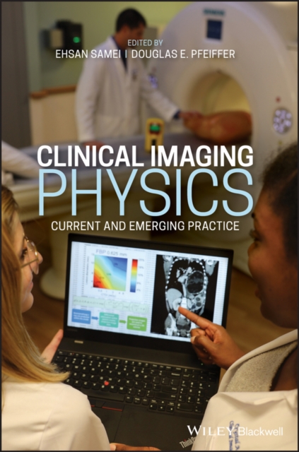
Clinical imaging physics : current and emerging practice
Clinical Medical Imaging Physics: Current and Emerging Practice is the first text of its kind--a comprehensive reference work covering all imaging modalities in use in clinical medicine today. Destined to become a classic in the field, this book provides state-of-practice descriptions for each imaging modality, followed by special sections on new and emerging applications, technologies, and practices.
Authored by luminaries in the field of medical physics, this resource is a sophisticated, one-volume handbook to a fast-advancing field that is becoming ever more central to contemporary clinical medicine.
* Summarizes the current state of clinical medical imaging physics in one volume, with a focus on emerging technologies and applications
* Provides comprehensive coverage of all key clinical imaging modalities, taking into account the new realities in healthcare practice
* Features a strong focus on clinical application of principles and technology, now and in the future
* Contains authoritative text compiled by world-renowned editors and contributors responsible for guiding the development of the field
Practicing radiologists and medical physicists will appreciate Clinical Medical Imaging Physics as a peerless everyday reference work. Additionally, graduate students and residents in medical physics and radiology will find this book essential as they study for their board exams.
eBook

Phase-contrast virtual chest radiography
Respiratory X-ray imaging enhanced by phase contrast has shown improved airway visualization in animal models. Limitations in current X-ray technology have nevertheless hindered clinical translation, leaving the potential clinical impact an open question. Here, we explore phase-contrast chest radiography in a realistic in silico framework. Specifically, we use preprocessed virtual patients to generate in silico chest radiographs by Fresnel-diffraction simulations of X-ray wave propagation. Following a reader study conducted with clinical radiologists, we predict that phase-contrast edge enhancement will have a negligible impact on improving solitary pulmonary nodule detection (6 to 20 mm). However, edge enhancement of bronchial walls visualizes small airways (<2 mm), which are invisible in conventional radiography. Our results show that phase-contrast chest radiography could play a future role in observing small-airway obstruction (e.g., relevant for asthma or early-stage chronic obstructive pulmonary disease), which cannot be directly visualized using current clinical methods, thereby motivating the experimental development needed for clinical translation. Finally, we discuss quantitative requirements on distances and X-ray source/detector specifications for clinical implementation of phase-contrast chest radiography.
Journal Article
How accurate and precise are CT based measurements of iodine concentration? A comparison of the minimum detectable concentration difference among single source and dual source dual energy CT in a phantom study
ObjectivesTo assess the impact of scan- and patient-related factors on the error and the minimum detectable difference in iodine concentration among different generations of single-source (SS) fast kV-switching and dual-source (DS) dual-energy CT (DECT).MethodsLesions having eight different iodine concentrations (0.2–4 mgI/mL) were emulated in a 3D-printed phantom of medium and large size. Each combination of concentration and size was scanned in dual-energy mode on four different SS and DS DECTs. Radiation doses were 7 and 10 mGy (medium size) and 10, 13, and 16 mGy (large size). Iodine maps were reconstructed with filtered back projection (FBP) and vendor-specific iterative reconstruction algorithms (IRs). Absolute error of iodine quantification (E) was measured. Multivariate regression models determined the influence of CT scanner, iodine concentration, phantom size, radiation dose, and reconstruction algorithm on E. The minimum detectable difference in iodine concentration (ICmin) under the same imaging conditions (intra-conditional) and among different imaging conditions (inter-conditional) was calculated.ResultsThe error was significantly lower in current than in previous DECT generations (p < 0.001). For all CT scanner conditions, the error was significantly higher with increasing phantom size and decreasing radiation dose (p < 0.001). Iodine concentration only significantly affected the error for SS DECT (p < 0.001). ICmin depended on patient- and scan-related factors and ranged from 0.4 to 1.5 mgI/mL.ConclusionsPatient- and scan-related factors have a significant impact on the error and minimum detectable difference in iodine concentration within and among SS fast kV-switching and DS DECT.Key Points• Patient- and scan-related factors have a significant impact on the error and minimum detectable difference in dual-energy CT-based iodine quantification.• Third-generation DECTs outperformed second-generation scanners for both single-source and dual-source dual-energy CT.• The minimum intra- and inter-conditional detectable difference in iodine concentration ranged from 0.4 to 1.5 mg iodine/mL.
Journal Article
Development of a deep learning based approach for multi-material decomposition in spectral CT: a proof of principle in silico study
Conventional approaches to material decomposition in spectral CT face challenges related to precise algorithm calibration across imaged conditions and low signal quality caused by variable object size and reduced dose. In this proof-of-principle study, a deep learning approach to multi-material decomposition was developed to quantify iodine, gadolinium, and calcium in spectral CT. A dual-phase network architecture was trained using synthetic datasets containing computational models of cylindrical and virtual patient phantoms. Classification and quantification performance was evaluated across a range of patient size and dose parameters. The model was found to accurately classify (accuracy: cylinders – 98%, virtual patients – 97%) and quantify materials (mean absolute percentage difference: cylinders – 8–10%, virtual patients – 10–15%) in both datasets. Performance in virtual patient phantoms improved as the hybrid training dataset included a larger contingent of virtual patient phantoms (accuracy: 48% with 0 virtual patients to 97% with 8 virtual patients). For both datasets, the algorithm was able to maintain strong performance under challenging conditions of large patient size and reduced dose. This study shows the validity of a deep-learning based approach to multi-material decomposition trained with in-silico images that can overcome the limitations of conventional material decomposition approaches.
Journal Article

Clinical concordance with Image Gently guidelines for pediatric computed tomography: a study across 663,417 CT scans at 53 clinical facilities
BackgroundManaging patient radiation dose in pediatric computed tomography (CT) examinations is essential. Some organizations, most notably Image Gently, have suggested techniques to lower dose to pediatric patients and mitigate risk while maintaining image quality.ObjectiveWe sought to validate whether institutions are observing Image Gently guidelines in practice.Materials and methodsDose-relevant data from 663,417 abdomen-pelvis and chest CT scans were obtained from 53 facilities. Patients were assigned arbitrary age cohorts with a minimum size of n=12 patients in each age group, for statistical purposes. All pediatric (<19 years old) cohorts at a given facility were compared to the adult cohort by a Kruskal-Wallis test for each of the four scan parameters — (1) x-ray tube kilovoltage (kV), (2) tube-current-by-exposure-time product (tube mAs), (3) scan pitch and (4) tube rotation time — to assess whether the distribution of values in the pediatric cohorts differed from the adult cohort. The same was repeated with volume CT dose index (CTDIvol) and size-specific dose estimate (SSDE) to assess whether pediatric cohorts received less dose than adult cohorts. A P-value of <0.05 was deemed significant.ResultsAcross the 150 pediatric cohorts, 134 had scan parameters that were more child-sized than their adult counterparts. In 128 of these 134 pediatric cohorts, the CTDIvol was less than the adult counterpart. In 111 of these 128 pediatric cohorts, the SSDE was less than the adult counterpart.ConclusionThe study reaffirms that in practice, Image Gently’s suggestions of lowering tube mAs and peak kilovoltage are commonly employed and effective at reducing pediatric CT dose.
Journal Article
Minimum perceivable size difference: how well can radiologists visually detect a change in lung nodule size from CT images?
Objective
The purpose of this study was to determine how well radiologists could visually detect a change in lung nodule size on the basis of visual image perception alone.
Subjects and methods
Under IRB approval, 109 standard chest CT image series were anonymized and exported from PACS. Nine hundred forty virtual lung nodule pairs (six baseline diameters, six relative volume differences, two nodule types—solid and ground glass—and 14 repeats) were digitally inserted into the chest CT image series (same location, different sizes between the pair). These digitally altered CT image pairs were shown to nine radiologists who were tasked to visually determine which image contained the larger nodule using a two-alternative forced-choice perception experimental design. These data were statistically analyzed using a generalized linear mixed effects model to determine how accurately the radiologists were able to correctly identify the larger nodule.
Results
Nominal baseline nodule diameter, relative volume difference, and nodule type were found to be statistically significant factors (
p
< 0.001) in influencing the radiologists’ accuracy. For solid (ground-glass) nodules, the baseline diameter needed to be at least 6.3 mm (13.2 mm) to be able to visually detect a 25% change in volume with 95 ± 1.4% accuracy. Accuracy was lowest for the nodules with the smallest baseline diameters and smallest relative volume differences. Additionally, accuracy was lower for ground-glass nodules compared to solid nodules.
Conclusions
Factors that impacted visual size assessment were baseline nodule diameter, relative volume difference, and solid versus non-solid nodule type, with larger and more solid lesions offering a more precise assessment of change.
Key Points
• For solid nodules, radiologists could visually detect a 25% change in volume with 95% accuracy for nodules having greater than 6.3-mm baseline diameter.
• For ground-glass nodules, radiologists could visually detect a 25% change in volume with 95% accuracy for nodules having greater than 13.2-mm baseline diameter.
• Accuracy in detecting a change in nodule size began to stabilize around 90–100% for nodules with larger baseline diameters (> 8 mm for solid nodules, > 12 mm for ground-glass nodules) and larger relative volume differences (>15% for solid nodules, > 25% for ground-glass nodules).
Journal Article
Correction to: Comparison of 12 surrogates to characterize CT radiation risk across a clinical population
A Correction to this paper has been published:
https://doi.org/10.1007/s00330-021-07903-z
Journal Article