Catalogue Search | MBRL


Are you sure you want to remove the book from the shelf?

{{itemTitle}}
988
result(s) for
"Zhao, Jean"
Sort by:

Targeting CDK4 and CDK6 in cancer
Cyclin-dependent kinase 4 (CDK4) and CDK6 are critical mediators of cellular transition into S phase and are important for the initiation, growth and survival of many cancer types. Pharmacological inhibitors of CDK4/6 have rapidly become a new standard of care for patients with advanced hormone receptor-positive breast cancer. As expected, CDK4/6 inhibitors arrest sensitive tumour cells in the G1 phase of the cell cycle. However, the effects of CDK4/6 inhibition are far more wide-reaching. New insights into their mechanisms of action have triggered identification of new therapeutic opportunities, including the development of novel combination regimens, expanded application to a broader range of cancers and use as supportive care to ameliorate the toxic effects of other therapies. Exploring these new opportunities in the clinic is an urgent priority, which in many cases has not been adequately addressed. Here, we provide a framework for conceptualizing the activity of CDK4/6 inhibitors in cancer and explain how this framework might shape the future clinical development of these agents. We also discuss the biological underpinnings of CDK4/6 inhibitor resistance, an increasingly common challenge in clinical oncology.Dysregulation of cyclin-dependent kinase 4 (CDK4) and CDK6, regulators of the cell cycle, favours the growth and survival of several cancer types. Owing to this, CDK4 and CDK6 inhibitors were developed and are currently approved for the treatment of advanced hormone receptor-positive breast cancer. This Review describes how we are only now beginning to fully understand their mechanisms of action and provides a new framework for conceptualizing their activity, which might enable expansion of the clinical opportunities of these agents.
Journal Article
PI3K in cancer: divergent roles of isoforms, modes of activation and therapeutic targeting
Key Points
Oncogenic mutation of the phosphatidylinositol 3-kinase (PI3K) catalytic isoform p110α is frequent in human cancers, whereas the catalytic isoforms p110β, p110δ and p110γ are rarely mutated but can be overexpressed. Mutation or loss of expression of regulatory isoform p85α is also associated with cancer.
Although class IA PI3K catalytic isoforms share structural and substrate similarities, they have specific roles in mediating PI3K signalling in different physiological and oncogenic contexts.
Cancer cells with upregulation or mutation of receptor tyrosine kinases (RTKs), oncogenic RAS mutations or activating p110α mutations are highly dependent on p110α, even in the presence of mutation or loss of PTEN.
In many cases, tumorigenesis that is driven by PTEN loss depends on p110β. However, PI3K isoform dependence in PTEN-deficient transformation may be governed by other PI3K isoforms that are dominant in a tissue or compartment, or shifted by coexisting oncogenic mutations.
Isoforms p110α, p110δ and p110γ bind to and are activated by RAS subfamily GTPases, while p110β binds to and is activated by RHO subfamily GTPases RAC1 and CDC42.
Non-isoform-selective pan-PI3K inhibitors have not yielded exciting clinical results, but second-generation PI3K drugs that target individual PI3K isoforms may be able to achieve greater therapeutic efficacy by offering improved specificity and reduced toxicity.
The p110δ-selective inhibitor idelalisib has been remarkably effective in clinical trials for patients with B cell malignancies, while p110α-selective inhibitors have shown promise in early-phase trials for patients with solid tumours with
PIK3CA
mutations or
HER2
amplification.
Intrinsic and acquired resistance mechanisms are a continuing challenge for PI3K-directed therapeutic approaches. To overcome this, combination therapies and alternative dosing strategies are being developed and evaluated in both preclinical and clinical settings.
Hyperactivation of phosphatidylinositol 3-kinase (PI3K) signalling cascades is one of the most common events in human cancers. This Review discusses recent advances in our knowledge of the roles of distinct PI3K isoforms in normal and oncogenic signalling, and the current state and future potential of targeting this pathway in the clinic.
Phosphatidylinositol 3-kinases (PI3Ks) are crucial coordinators of intracellular signalling in response to extracellular stimuli. Hyperactivation of PI3K signalling cascades is one of the most common events in human cancers. In this Review, we discuss recent advances in our knowledge of the roles of specific PI3K isoforms in normal and oncogenic signalling, the different ways in which PI3K can be upregulated, and the current state and future potential of targeting this pathway in the clinic.
Journal Article
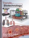
Bioprinting for cancer research
•Bioprinting techniques can be used to create 3D tumor models for cancer research.•Bioprinting methods include inkjet, microextrusion, and laser technologies.•3D cancer models mimic the tumor microenvironment better than 2D models.•This will lead to a better understanding of cancer and aid in cancer drug screening.
Bioprinting offers the ability to create highly complex 3D architectures with living cells. This cutting-edge technique has significantly gained popularity and applicability in several fields. Bioprinting methods have been developed to effectively and rapidly pattern living cells, biological macromolecules, and biomaterials. These technologies hold great potential for applications in cancer research. Bioprinted cancer models represent a significant improvement over previous 2D models by mimicking 3D complexity and facilitating physiologically relevant cell–cell and cell–matrix interactions. Here we review bioprinting methods based on inkjet, microextrusion, and laser technologies and compare 3D cancer models with 2D cancer models. We discuss bioprinted models that mimic the tumor microenvironment, providing a platform for deeper understanding of cancer pathology, anticancer drug screening, and cancer treatment development.
Journal Article

Lactate regulates cell cycle by remodelling the anaphase promoting complex
Lactate is abundant in rapidly dividing cells owing to the requirement for elevated glucose catabolism to support proliferation
1
–
6
. However, it is not known whether accumulated lactate affects the proliferative state. Here we use a systematic approach to determine lactate-dependent regulation of proteins across the human proteome. From these data, we identify a mechanism of cell cycle regulation whereby accumulated lactate remodels the anaphase promoting complex (APC/C). Remodelling of APC/C in this way is caused by direct inhibition of the SUMO protease SENP1 by lactate. We find that accumulated lactate binds and inhibits SENP1 by forming a complex with zinc in the SENP1 active site. SENP1 inhibition by lactate stabilizes SUMOylation of two residues on APC4, which drives UBE2C binding to APC/C. This direct regulation of APC/C by lactate stimulates timed degradation of cell cycle proteins, and efficient mitotic exit in proliferative human cells. This mechanism is initiated upon mitotic entry when lactate abundance reaches its apex. In this way, accumulation of lactate communicates the consequences of a nutrient-replete growth phase to stimulate timed opening of APC/C, cell division and proliferation. Conversely, persistent accumulation of lactate drives aberrant APC/C remodelling and can overcome anti-mitotic pharmacology via mitotic slippage. In sum, we define a biochemical mechanism through which lactate directly regulates protein function to control the cell cycle and proliferation.
Discovery of a biochemical mechanism through which lactate binds and inhibits the SUMO protease SENP1, stimulating timed degradation of cell cycle proteins, and resulting in mitotic exit.
Journal Article

Targeting the phosphoinositide 3-kinase pathway in cancer
Key Points
The phosphoinositide 3-kinase (PI3K) pathway is a crucial signal transduction system linking the activation of receptor tyrosine kinases (RTKs), G protein-coupled receptors (GPCRs) and oncogenes such as
RAS
to multiple essential cellular functions.
The PI3K pathway is tightly controlled by a class of PI3Ks that generate the lipid second messenger phosphatidylinositol-3,4,5-trisphosphate (PtdIns(3,4,5)P
3
), and the tumor suppressor PTEN (phosphatase and tensin homologue), a lipid phosphatase that dephosphorylates PtdIns(3,4,5)P
3
, thereby counteracting the actions of PI3Ks.
The PI3K pathway is one of the most frequently activated signalling pathways in human cancer. Oncogenic activation of this pathway commonly occurs through activating mutations in the p110α isoform of PI3K or through loss of the PTEN tumour suppressor.
Inhibitors that target key components of this pathway, including PI3K, AKT and mammalian target of rapamycin (mTOR), are being actively developed. Some of them have reached clinical trials in patients with various solid and haematological malignancies. Most PI3K inhibitors developed to date are pan-PI3K inhibitors.
It may be desirable to generate isoform-specific PI3K inhibitors as PI3Ks have essential roles in a wide range of normal physiological functions, including glucose homeostasis and immune responses. For example, by targeting p110α or p110β isoforms in solid tumours, potential drugs might avoid toxicity to the immune system, which is largely dependent on p110δ and p110γ for function.
The presence of multiple nodes with feedback loops and crosstalk between pathways may affect therapeutic outcomes. Emerging strategies, such as simultaneously targeting two kinases in the pathway or the combination of PI3K pathway inhibitors with drugs that target other pathways, may achieve optimal clinical benefits.
The phosphoinositide 3-kinase (PI3K) signal transduction pathway is often inappropriately activated in human cancer and is crucial for cancer cell growth and survival. Liu and colleagues discuss key therapeutic targets in the PI3K pathway and agents that are currently in development, and consider the associated challenges and possible approaches to overcome them.
The phosphoinositide 3-kinase (PI3K) pathway is a key signal transduction system that links oncogenes and multiple receptor classes to many essential cellular functions, and is perhaps the most commonly activated signalling pathway in human cancer. This pathway therefore presents both an opportunity and a challenge for cancer therapy. Even as inhibitors that target PI3K isoforms and other major nodes in the pathway, including AKT and mammalian target of rapamycin (mTOR), reach clinical trials, major issues remain. Here, we highlight recent progress that has been made in our understanding of the PI3K pathway and discuss the potential of and challenges for the development of therapeutic agents that target this pathway in cancer.
Journal Article

STING agonism reprograms tumor-associated macrophages and overcomes resistance to PARP inhibition in BRCA1-deficient models of breast cancer
PARP inhibitors (PARPi) have drastically changed the treatment landscape of advanced ovarian tumors with
BRCA
mutations. However, the impact of this class of inhibitors in patients with advanced
BRCA
-mutant breast cancer is relatively modest. Using a syngeneic genetically-engineered mouse model of breast tumor driven by
Brca1
deficiency, we show that tumor-associated macrophages (TAMs) blunt PARPi efficacy both in vivo and in vitro. Mechanistically, BRCA1-deficient breast tumor cells induce pro-tumor polarization of TAMs, which in turn suppress PARPi-elicited DNA damage in tumor cells, leading to reduced production of dsDNA fragments and synthetic lethality, hence impairing STING-dependent anti-tumor immunity. STING agonists reprogram M2-like pro-tumor macrophages into an M1-like anti-tumor state in a macrophage STING-dependent manner. Systemic administration of a STING agonist breaches multiple layers of tumor cell-mediated suppression of immune cells, and synergizes with PARPi to suppress tumor growth. The therapeutic benefits of this combination require host STING and are mediated by a type I IFN response and CD8
+
T cells, but do not rely on tumor cell-intrinsic STING. Our data illustrate the importance of targeting innate immune suppression to facilitate PARPi-mediated engagement of anti-tumor immunity in breast cancer.
PARP inhibitor (PARPi) therapy has demonstrated only modest efficacy in advanced breast cancer with BRCA mutations. Here the authors show that, by suppressing PARPi-triggered DNA damage and reducing dsDNA production in BRCA1-deficient breast tumor cells, tumor associated macrophages contribute to PARPi resistance, that can be overcome by STING agonism.
Journal Article

Statin-dye conjugates for selective targeting of KRAS mutant cancer cells
Over 90% of pancreatic ductal adenocarcinoma (PDAC) patients involve KRAS mutations ( KRAS MUT ), for which current treatment options are limited. Statins, commonly used to lower cholesterol, have demonstrated certain selective toxicity towards KRAS -transformed cells, prompting the question of whether statin-based conjugates could achieve selective uptake specifically in KRAS MUT cells. To investigate this, we synthesized statin-dye conjugates by attaching a fluorescent dye (Cy5.5) to two statins: simvastatin and pravastatin, aiming to assess whether selective uptake indeed occurs. Our findings revealed that these conjugates exhibited markedly enhanced uptake in KRAS MUT cells compared to KRAS wild-type ( KRAS WT ) cells. We evaluated the uptake of these conjugates in both KRAS MUT and KRAS WT cells and examined their potential to selectively target KRAS MUT pancreatic cancer cells (PCCs) using an engineered PDAC tumor model co-cultured with PCCs and cancer-associated fibroblasts (CAFs). Our findings indicate that KRAS MUT cancer cells exhibited higher uptake of statin-Cy5.5 conjugates via enhanced macropinocytosis compared to KRAS WT cancer cells and CAFs. We also found enhanced uptake of the statin-Cy5.5 conjugate in MCF10A cells with PTEN deficiency, a condition known to elevate macropinocytosis, compared to control MCF10A cells with wild-type PTEN . Notably, in the PCC and CAF co-culture model, the pravastatin-Cy5.5 conjugate selectively killed KRAS MUT PCCs without affecting the KRAS WT CAFs. These findings highlight the unique synergistic potential of statin-Cy5.5—distinct from either component alone—as targeted delivery vehicles for KRAS MUT cancer therapy.
Journal Article
Targeting neuronal activity-regulated neuroligin-3 dependency in high-grade glioma
The growth of adult and paediatric brain tumours depends on a microenvironmental signalling pathway involving the activity-regulated secretion of neuroligin-3 (NLGN3) from normal neurons and oligodendrocyte precursor cells, highlighting the potential of NLGN3 as a therapeutic target.
A roadblock to brain tumours
The growth of brain tumours is influenced by the microenvironment of the normal brain. Here, the authors unravel a microenvironmental signalling pathway involving secretion of neuroligin-3 (NLGN3) by normal neurons. NLGN3 is cleaved by extracellular proteases, stimulates oncogenic signalling in glioma cells and induces transcriptional changes. Inhibition of NLGN3 cleavage in the tumour microenvironment impairs the growth of brain tumour xenografts
in vivo
, suggesting that interfering with this pathway and with microenvironmental signalling in general is a potential therapeutic strategy for brain tumours.
High-grade gliomas (HGG) are a devastating group of cancers, and represent the leading cause of brain tumour-related death in both children and adults. Therapies aimed at mechanisms intrinsic to glioma cells have translated to only limited success; effective therapeutic strategies will need also to target elements of the tumour microenvironment that promote glioma progression. Neuronal activity promotes the growth of a range of molecularly and clinically distinct HGG types, including adult and paediatric glioblastoma (GBM), anaplastic oligodendroglioma, and diffuse intrinsic pontine glioma (DIPG)
1
. An important mechanism that mediates this neural regulation of brain cancer is activity-dependent cleavage and secretion of the synaptic adhesion molecule neuroligin-3 (NLGN3), which promotes glioma proliferation through the PI3K–mTOR pathway
1
. However, the necessity of NLGN3 for glioma growth, the proteolytic mechanism of NLGN3 secretion, and the further molecular consequences of NLGN3 secretion in glioma cells remain unknown. Here we show that HGG growth depends on microenvironmental NLGN3, identify signalling cascades downstream of NLGN3 binding in glioma, and determine a therapeutically targetable mechanism of secretion. Patient-derived orthotopic xenografts of paediatric GBM, DIPG and adult GBM fail to grow in
Nlgn3
knockout mice. NLGN3 stimulates several oncogenic pathways, such as early focal adhesion kinase activation upstream of PI3K–mTOR, and induces transcriptional changes that include upregulation of several synapse-related genes in glioma cells. NLGN3 is cleaved from both neurons and oligodendrocyte precursor cells via the ADAM10 sheddase. ADAM10 inhibitors prevent the release of NLGN3 into the tumour microenvironment and robustly block HGG xenograft growth. This work defines a promising strategy for targeting NLGN3 secretion, which could prove transformative for HGG therapy.
Journal Article

Targeting tumor-associated macrophages with STING agonism improves the antitumor efficacy of osimertinib in a mouse model of EGFR-mutant lung cancer
Despite the impressive clinical response rate of osimertinib, a third-generation EGFR-TKI, as a frontline treatment for patients with EGFR-mutant non-small-cell lung cancer (NSCLC) or as a salvage therapy for patients with T790M mutation, resistance to osimertinib is common in the clinic. The mechanisms underlying osimertinib resistance are heterogenous. While genetic mutations within EGFR or other cancer driver pathways mediated mechanisms are well-documented, the role of tumor cell and tumor immune microenvironment in mediating the response to osimertinib remains elusive.
Here, using a syngeneic mouse model of EGFR-mutant lung cancer, we show that tumor regression elicited by osimertinib requires activation of CD8+ T cells. However, tumor-associated macrophages (TAMs) accumulated in advanced tumors inhibit CD8+ T cell activation and diminish the response to osimertinib. These results are corroborated by analyses of clinical data. Notably, reprogramming TAMs with a systemic STING agonist MSA-2 reinvigorates antitumor immunity and leads to durable tumor regression in mice when combined with osimertinib.
Our results reveal a new mechanism of EGFR-TKI resistance and suggest a new therapeutic strategy for the treatment of EGFR-mutant tumors.
Journal Article
CDK4/6 inhibition triggers anti-tumour immunity
Mouse models of breast carcinoma and other solid tumours show that selective cyclin-dependent kinase 4 and 6 (CDK4/6) inhibitors not only induce tumour cell cycle arrest but also promote anti-tumour immunity.
Anti-tumour effect of CDK4/6 inhibitors
CDK4/6 inhibitors are currently in clinical trials for breast cancer. They are known to induce cytostatic effects. Here, the authors uncover a novel effect of CDK4/6 inhibitors that could contribute to their therapeutic effect. By both increasing tumour cell antigen presentation and suppressing the proliferation of regulatory T cells, CDK4/6 inhibitors enhance anti-tumour immunity. These effects are each linked to reduced expression of DNMT1 after treatment.
In vivo
, combining CDK4/6 inhibitors and checkpoint blockade provides a more profound anti-tumour effect.
Cyclin-dependent kinases 4 and 6 (CDK4/6) are fundamental drivers of the cell cycle and are required for the initiation and progression of various malignancies
1
,
2
. Pharmacological inhibitors of CDK4/6 have shown significant activity against several solid tumours
3
,
4
. Their primary mechanism of action is thought to be the inhibition of phosphorylation of the retinoblastoma tumour suppressor, inducing G1 cell cycle arrest in tumour cells
5
. Here we use mouse models of breast carcinoma and other solid tumours to show that selective CDK4/6 inhibitors not only induce tumour cell cycle arrest, but also promote anti-tumour immunity. We confirm this phenomenon through transcriptomic analysis of serial biopsies from a clinical trial of CDK4/6 inhibitor treatment for breast cancer. The enhanced anti-tumour immune response has two underpinnings. First, CDK4/6 inhibitors activate tumour cell expression of endogenous retroviral elements, thus increasing intracellular levels of double-stranded RNA. This in turn stimulates production of type III interferons and hence enhances tumour antigen presentation. Second, CDK4/6 inhibitors markedly suppress the proliferation of regulatory T cells. Mechanistically, the effects of CDK4/6 inhibitors both on tumour cells and on regulatory T cells are associated with reduced activity of the E2F target, DNA methyltransferase 1. Ultimately, these events promote cytotoxic T-cell-mediated clearance of tumour cells, which is further enhanced by the addition of immune checkpoint blockade. Our findings indicate that CDK4/6 inhibitors increase tumour immunogenicity and provide a rationale for new combination regimens comprising CDK4/6 inhibitors and immunotherapies as anti-cancer treatment.
Journal Article