
Catalogue Search | MBRL


Are you sure you want to remove the book from the shelf?

{{itemTitle}}
34,933
result(s) for
"Brain aging"
Sort by:

Successful aging : a neuroscientist explores the power and potential of our lives
\"Author of the iconic bestsellers This Is Your Brain on Music and The Organized Mind, Daniel Levitin turns his keen insights to what happens in our brains as we age; why we should think about health span, not life span; and, based on a rigorous analysis of neuroscientific evidence, how you can make the most of your seventies, eighties, and nineties today, no matter how old you are now\"-- Provided by publisher.
Microglial cathepsin B as a key driver of inflammatory brain diseases and brain aging
Interleukin-1β is a potent proinflammatory cytokine that plays a key role in the pathogenesis of the brain aging and diverse range of neurological diseases including Alzheimer's disease, Parkinson's disease, stroke and persistent pain. Activated microglia are the main cellular source of interleukin-1β in the brain. Cathepsin B is associated with the production and secretion of interleukin-1β through pyrin domain-containing protein 3 inflammasome-independent processing of procaspase-3 in the phagolysosomes. The leakage of cathepsin B from the endosomal-lysosomal system during aging is associated with the proteolytic degradation of mitochondrial transcription factor A, which can stabilize mitochondrial DNA. Therefore, microglial cathepsin B could function as a major driver for inflammatory brain diseases and brain aging. Orally active and blood-brain barrier-permeable specific inhibitors for cathepsin B can be potentially effective new pharmaceutical interventions against inflammatory brain diseases and brain aging.
Journal Article

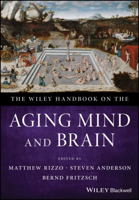
The Wiley Handbook on the Aging Mind and Brain
A thought-provoking treatise on understanding and treating the aging mind and brain This handbook recognizes the critical issues surrounding mind and brain health by tackling overarching and pragmatic needs so as to better understand these multifaceted issues. This includes summarizing and synthesizing critical evidence, approaches, and strategies from multidisciplinary research—all of which have advanced our understanding of the neural substrates of attention, perception, memory, language, decision-making, motor behavior, social cognition, emotion, and other mental functions. Written by a plethora of health experts from around the world, The Wiley Handbook on the Aging Mind and Brain offers in-depth contributions in 7 sections: Introduction; Methods of Assessment; Brain Functions and Behavior across the Lifespan; Cognition, Behavior and Disease; Optimizing Brain Function in Health and Disease; Forensics, Competence, Legal, Ethics and Policy Issues; and Conclusion and New Directions. * Geared toward improving the recognition, diagnosis, and treatment of many brain-based disorders that occur in older adults and that cause disability and death * Seeks to advance the care of patients who have perceptual, cognitive, language, memory, emotional, and many other behavioral symptoms associated with these disorders * Addresses principles and practice relevant to challenges posed by the US National Academy of Sciences and National Institute of Aging (NIA) * Presents materials at a scientific level that is appropriate for a wide variety of providers The Wiley Handbook on the Aging Mind and Brain is an important text for neurologists, psychiatrists, psychologists, physiatrists, geriatricians, nurses, pharmacists, social workers, and other primary caregivers who care for patients in routine and specialty practices as well as students, interns, residents, and fellows.
eBook

The aging brain : functional adaptation across adulthood
\"Brain aging has long been seen as a process of deterioration and decline. Today, this view been challenged with research showing that not all cognitive processes decline with age, that some improve over the course of adulthood, and those that improve can often compensate for those that decline. Chapters in this multidisciplinary volume examine the neural mechanisms underlying changes in the aging brain, changes in learning and memory, risk and protective factors, and the assessment and prevention of cognitive decline\"--Provided by publisher.
Book

Fast three‐dimensional image generation for healthy brain aging using diffeomorphic registration
Predicting brain aging can help in the early detection and prognosis of neurodegenerative diseases. Longitudinal cohorts of healthy subjects scanned through magnetic resonance imaging (MRI) have been essential to understand the structural brain changes due to aging. However, these cohorts suffer from missing data due to logistic issues in the recruitment of subjects. This paper proposes a methodology for filling up missing data in longitudinal cohorts with anatomically plausible images that capture the subject‐specific aging process. The proposed methodology is developed within the framework of diffeomorphic registration. First, two novel modules are introduced within Synthmorph, a fast, state‐of‐the‐art deep learning‐based diffeomorphic registration method, to simulate the aging process between the first and last available MRI scan for each subject in three‐dimensional (3D). The use of image registration also makes the generated images plausible by construction. Second, we used six image similarity measurements to rearrange the generated images to the specific age range. Finally, we estimated the age of every generated image by using the assumption of linear brain decay in healthy subjects. The methodology was evaluated on 2662 T1‐weighted MRI scans from 796 healthy participants from 3 different longitudinal cohorts: Alzheimer's Disease Neuroimaging Initiative, Open Access Series of Imaging Studies‐3, and Group of Neuropsychological Studies of the Canary Islands (GENIC). In total, we generated 7548 images to simulate the access of a scan per subject every 6 months in these cohorts. We evaluated the quality of the synthetic images using six quantitative measurements and a qualitative assessment by an experienced neuroradiologist with state‐of‐the‐art results. The assumption of linear brain decay was accurate in these cohorts (R2 ∈ [.924, .940]). The experimental results show that the proposed methodology can produce anatomically plausible aging predictions that can be used to enhance longitudinal datasets. Compared to deep learning‐based generative methods, diffeomorphic registration is more likely to preserve the anatomy of the different structures of the brain, which makes it more appropriate for its use in clinical applications. The proposed methodology is able to efficiently simulate anatomically plausible 3D MRI scans of brain aging of healthy subjects from two images scanned at two different time points. In this work, we proposed a methodology with the aim of simulating subject‐specific aging in brain magnetic resonance imaging (MRI) given two three‐dimensional images acquired at different time points. Deep learning‐based diffeomorphic registration was used as a backbone to generate deformation fields at different integration points. Similarity measurements were used for controlling the age estimation of the generated images by using a linear assumption.
Journal Article


Clinical value of structural cranial magnetic resonance imaging analysis in assessing brain aging in ischemic stroke patients
Objective
Using structural cranial magnetic resonance imaging (MRI), we constructed a structural Brain Aging Index (sBAI) and evaluated the brain aging status, expressed as Brain Predicted Age Difference (Brain-PAD), in acute ischemic stroke patients. We aim to investigate its associations with baseline clinical and imaging characteristics, small vessel disease (SVD) burden, and 12-month functional and cognitive outcomes, and explored its potential contribution to prognostic models.
Methods
A retrospective cohort of 150 patients with acute ischemic stroke admitted between November 2021 and November 2023 was analyzed. Diffusion-weighted imaging (DWI) infarct volume, white matter hyperintensity (WMH) volume, lacunes, cerebral microbleeds (CMBs), enlarged perivascular spaces (EPVS), hippocampal volume, and cortical thickness were collected. Clinical and imaging differences were compared between the “age-matched group” (Brain-PAD < + 5 years) and the “accelerated brain aging group” (Brain-PAD ≥ + 5 years).
Results
Patients in the accelerated brain aging group had significantly higher admission National Institutes of Health Stroke Scale (NIHSS) scores, larger DWI infarct volumes, greater WMH burden, and higher total SVD scores. During follow-up, the accelerated brain aging group had a lower rate of favorable functional outcome at discharge and a higher incidence of poor 12-month functional outcome, together with significantly lower MoCA scores. Admission NIHSS and WMH volume (aOR = 1.10) were independent predictors of 12-month functional outcome. sBAI was significantly associated with 12-month MoCA scores.
Conclusion
Brain aging status derived from structural MRI is closely associated with acute lesion burden, cumulative small vessel disease, and poststroke cognitive and functional outcomes.
Journal Article


Transcriptomic Changes Highly Similar to Alzheimer’s Disease Are Observed in a Subpopulation of Individuals During Normal Brain Aging
Aging is a major risk factor for late-onset Alzheimer’s disease (LOAD). How aging contributes to the development of LOAD remains elusive. In this study, we examined multiple large-scale transcriptomic datasets from both normal aging and LOAD brains to understand the molecular interconnection between aging and LOAD. We found that shared gene expression changes between aging and LOAD are mostly seen in the hippocampal and several cortical regions. In the hippocampus, the expression of phosphoprotein, alternative splicing and cytoskeleton genes are commonly changed in both aging and AD, while synapse, ion transport, and synaptic vesicle genes are commonly down-regulated. Aging-specific changes are associated with acetylation and methylation, while LOAD-specific changes are more related to glycoprotein (both up- and down-regulations), inflammatory response (up-regulation), myelin sheath and lipoprotein (down-regulation). We also found that normal aging brain transcriptomes from relatively young donors (45–70 years old) clustered into several subgroups and some subgroups showed gene expression changes highly similar to those seen in LOAD brains. Using brain transcriptomic datasets from another cohort of older individuals (>70 years), we found that samples from cognitively normal older individuals clustered with the “healthy aging” subgroup while AD samples mainly clustered with the “AD similar” subgroups. This may imply that individuals in the healthy aging subgroup will likely remain cognitively normal when they become older and vice versa. In summary, our results suggest that on the transcriptome level, aging and LOAD have strong interconnections in some brain regions in a subpopulation of cognitively normal aging individuals. This supports the theory that the initiation of LOAD occurs decades earlier than the manifestation of clinical phenotype and it may be essential to closely study the “normal brain aging” to identify the very early molecular events that may lead to LOAD development.
Journal Article