Catalogue Search | MBRL


Are you sure you want to remove the book from the shelf?

{{itemTitle}}
115,663
result(s) for
"Cardiovascular imaging"
Sort by:

Cardiac T2 mapping: robustness and homogeneity of standardized in-line analysis
Background and purpose
Interpretation of T2 values remains difficult due to limited comparability across hardware and software systems and the lack of validated measurement recommendations for the number and orientation of mandatory slices. Our aims were to provide a standardized comparison of intra- and inter-individual T2 values in the short and long axes and to investigate inter-scanner reproducibility.
Method and materials
Ninety cardiovascular magnetic resonance (CMR) studies in 30 healthy subjects were performed with three identical 1.5 T CMR scanners (same hardware and software) using a balanced steady-state free precession (bSSFP) gradient echo sequence in three short axis (SAx) and three long axis (LAx) views. A commercially available T2 mapping software package of the latest generation with automatic in-line motion correction was used for acquisition. Regions of interest were manually drawn in each of the 16 myocardial segments according to the American Heart Association (AHA) model in three SAx and three LAx acquisitions. Analysis of inter-scanner, inter-segmental, intra-segmental, inter-regional and inter-level differences was performed.
Results
Inter-scanner reproducibility was high and the mean myocardial T2 value for all evaluated segments was 45.7 ± 3.4 ms. Significant inter-segmental variations of mean T2 values were found. Mean intra-segmental T2 values were comparable between LAx and SAx acquisitions in 72%. Significantly higher T2 values were found in apical segments and a significant disparity among different regions was found for SAx and LAx orientations.
Conclusion
Standardized cardiac T2 mapping is highly reproducible on identical CMR systems. T2 values vary significantly between single heart segments, regions, levels, and axes in young, healthy subjects.
Journal Article

Machine Learning in Cardiovascular Imaging: A Scoping Review of Published Literature
Purpose of Review
In this study, we planned and carried out a scoping review of the literature to learn how machine learning (ML) has been investigated in cardiovascular imaging (CVI).
Recent Findings
During our search, we found numerous studies that developed or utilized existing ML models for segmentation, classification, object detection, generation, and regression applications involving cardiovascular imaging data. We first quantitatively investigated the different aspects of study characteristics, data handling, model development, and performance evaluation in all studies that were included in our review. We then supplemented these findings with a qualitative synthesis to highlight the common themes in the studied literature and provided recommendations to pave the way for upcoming research.
Summary
ML is a subfield of artificial intelligence (AI) that enables computers to learn human-like decision-making from data. Due to its novel applications, ML is gaining more and more attention from researchers in the healthcare industry. Cardiovascular imaging is an active area of research in medical imaging with lots of room for incorporating new technologies, like ML.
Journal Article

CardiOvascular examination in awake Orangutans (Pongo pygmaeus pygmaeus): Low-stress Echocardiography including Speckle Tracking imaging (the COOLEST method)
Heart morphology as well as global and regional myocardial function can be assessed in awake orangutans with good to excellent repeatability and reproducibility. Conclusions This non-stressful method may be used for longitudinal cardiac follow-up in awake orangutans.
Journal Article

A population-based phenome-wide association study of cardiac and aortic structure and function
Differences in cardiac and aortic structure and function are associated with cardiovascular diseases and a wide range of other types of disease. Here we analyzed cardiovascular magnetic resonance images from a population-based study, the UK Biobank, using an automated machine-learning-based analysis pipeline. We report a comprehensive range of structural and functional phenotypes for the heart and aorta across 26,893 participants, and explore how these phenotypes vary according to sex, age and major cardiovascular risk factors. We extended this analysis with a phenome-wide association study, in which we tested for correlations of a wide range of non-imaging phenotypes of the participants with imaging phenotypes. We further explored the associations of imaging phenotypes with early-life factors, mental health and cognitive function using both observational analysis and Mendelian randomization. Our study illustrates how population-based cardiac and aortic imaging phenotypes can be used to better define cardiovascular disease risks as well as heart–brain health interactions, highlighting new opportunities for studying disease mechanisms and developing image-based biomarkers.
Using magnetic resonance images of the heart and aorta from 26,893 individuals in the UK Biobank, a phenome-wide association study associates cardiovascular imaging phenotypes with a wide range of demographic, lifestyle and clinical features.
Journal Article

Application of postmortem imaging modalities in cases of sudden death due to cardiovascular diseases–current achievements and limitations from a pathology perspective
Abstract Postmortem imaging (PMI) is increasingly used in postmortem practice and is considered a potential alternative to a conventional autopsy, particularly in case of sudden cardiac deaths (SCD). In 2017, the Association for European Cardiovascular Pathology (AECVP) published guidelines on how to perform an autopsy in such cases, which is still considered the gold standard, but the diagnostic value of PMI herein was not analyzed in detail. At present, significant progress has been made in the PMI diagnosis of acute ischemic heart disease, the most important cause of SCD, while the introduction of postmortem CT angiography (PMCTA) has improved the visualization of several parameters of coronary artery pathology that can support a diagnosis of SCD. Postmortem magnetic resonance (PMMR) allows the detection of acute myocardial injury-related edema. However, PMI has limitations when compared to clinical imaging, which severely impacts the postmortem diagnosis of myocardial injuries (ischemic versus non-ischemic), the age-dating of coronary occlusion (acute versus old), other potentially SCD-related cardiac lesions (e.g., the distinctive morphologies of cardiomyopathies), aortic diseases underlying dissection or rupture, or pulmonary embolism. In these instances, PMI cannot replace a histopathological examination for a final diagnosis. Emerging minimally invasive techniques at PMI such as image-guided biopsies of the myocardium or the aorta, provide promising results that warrant further investigations. The rapid developments in the field of postmortem imaging imply that the diagnosis of sudden death due to cardiovascular diseases will soon require detailed knowledge of both postmortem radiology and of pathology.
Journal Article

Coronary CT Angiography-Derived Fractional Flow Reserve in Asia and the United States: 2025 Status Update
Coronary CT-derived fractional flow reserve (CT-FFR) is a noninvasive alternative to invasive FFR for assessing the hemodynamic significance of coronary artery stenosis. CT-FFR uses routinely acquired coronary CT angiography (CCTA) with artificial intelligence and computational fluid dynamics to estimate pressure gradients, thereby supporting clinical decision-making without procedural risk. Adoption and implementation vary widely across regions. In Asia, South Korea exclusively uses HeartMedi+, which enables rapid analysis, streamlined workflow integration, and high user satisfaction. Japan relies primarily on FFR
, which has national reimbursement and significantly influences treatment strategies, reducing unnecessary invasive procedures. Hong Kong, Singapore, Taiwan, Thailand, and Vietnam remain in early or exploratory phases due to cost, reimbursement, and infrastructure barriers. In the United States, CT-FFR is guideline-endorsed and reimbursed, where HeartFlow is the most widely used, supported by robust clinical validation and outcome data. Despite regional variability, CT-FFR offers a noninvasive solution that enhances patient management, reduces unnecessary invasive testing, and is poised for broader clinical integration as technology and validation advance.
Journal Article

Relationship between right and left ventricle function in subjects free of cardiovascular diseases: a population-based MRI study
Right (RV) and left ventricular (LV) volumetric measurements by cardiac magnetic resonance imaging (MRI) are established for assessing systolic and diastolic function, but the role of MRI-derived lung volumes in LV function remains unclear. This study investigated the relationship between RV and LV function, considering lung volumes. In the KORA-MRI cohort, 361 subjects underwent 3 T whole-body MRI. Cardiac functional parameters were measured from cine-steady-state free precession sequences using cvi42. Lung volumes were derived semi-automatically with an in-house algorithm. Linear regression analyses assessed RV-LV relationships, adjusted for age, sex, cardiovascular risk factors, and lung volumes. Among 361 subjects (mean age 56.1 ± 9.1 years; 43% women), RV end-diastolic volume was positively associated with LV end-diastolic (β = 28.1, p < 0.001), end-systolic (β = 11.0, p < 0.001), and stroke volume (β = 17.0, p < 0.001), but inversely with ejection fraction (β = -1.4, p = 0.001). RV end-systole was positively associated with LV end-diastolic (β = 21.2, p < 0.001), end-systolic (β = 11.5, p < 0.001), stroke volume (β = 9.7, p < 0.001), and inversely with ejection fraction (β = -3.3, p < 0.001). Adjusting for lung volumes did not alter RV-LV associations, and no effect modification by sex was observed despite lung volume differences. In individuals without cardiovascular disease, RV and LV volumetric parameters were strongly associated, supporting the critical role of RV function in LV function, independent of lung volumes.
Journal Article
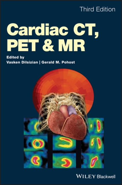
Cardiac CT, PET and MR
A complete guide to non-invasive imaging techniques in cardiology Today?s imaging technologies offer cardiologists more ways than ever to diagnose conditions of the heart without the need of endoscopies and other invasive procedures. Now in its third edition,Cardiac CT, PET and MRI continues to provide an in-depth explanation of these tools and their correct applications, while also exploring cardiac imaging?s most recent and groundbreaking developments. This wide-ranging guide places CT, PET and MRI in a practical context, illustrating clearly their respective functions as they apply to specific cardiological disorders and clinical situations. With the addition of seven new chapters, it also offers an expanded insight into PET – an increasingly popular and affordable diagnostic utility, hitherto underexplored in texts devoted to imaging. Cardiac CT, PET and MRI includes: * Clinically focused examinations of CT, PET and MRI – the three most popular non-invasive imaging modalities * Illustrative full-color photos and images * Access to a companion website featuring additional content Cardiologists, radiologists, nuclear medicine physicians, physicists, and imaging technologists alike will find the third edition of Cardiac CT, PET and MRI an informative and accessible resource with a direct use in their day-to-day practice.
eBook

Hybrid cardiovascular imaging. A clinical consensus statement of the european association of nuclear medicine (EANM) and the european association of cardiovascular imaging (EACVI) of the ESC
Hybrid imaging consists of a combination of two or more imaging modalities, which equally contribute to image information. To date, hybrid cardiovascular imaging can be performed by either merging images acquired on different scanners, or with truly hybrid PET/CT and PET/MR scanners. The European Association of Nuclear Medicine (EANM), and the European Association of Cardiovascular Imaging (EACVI) of the European Society of Cardiology (ESC) aim to review clinical situations that may benefit from the use of hybrid cardiac imaging and provide advice on acquisition protocols providing the most relevant information to reach diagnosis in various clinical situations.
Journal Article

Three-Dimensional Analysis of Vascular Development in the Mouse Embryo
Key vasculogenic (de-novo vessel forming) and angiogenic (vessel remodelling) events occur in the mouse embryo between embryonic days (E) 8.0 and 10.0 of gestation, during which time the vasculature develops from a simple circulatory loop into a complex, fine structured, three-dimensional organ. Interpretation of vascular phenotypes exhibited by signalling pathway mutants has historically been hindered by an inability to comprehensively image the normal sequence of events that shape the basic architecture of the early mouse vascular system. We have employed Optical Projection Tomography (OPT) using frequency distance relationship (FDR)-based deconvolution to image embryos immunostained with the endothelial specific marker PECAM-1 to create a high resolution, three-dimensional atlas of mouse vascular development between E8.0 and E10.0 (5 to 30 somites). Analysis of the atlas has provided significant new information regarding normal development of intersomitic vessels, the perineural vascular plexus, the cephalic plexus and vessels connecting the embryonic and extraembryonic circulation. We describe examples of vascular remodelling that provide new insight into the mechanisms of sprouting angiogenesis, vascular guidance cues and artery/vein identity that directly relate to phenotypes observed in mouse mutants affecting vascular development between E8.0 and E10.0. This atlas is freely available at http://www.mouseimaging.ca/research/mouse_atlas.html and will serve as a platform to provide insight into normal and abnormal vascular development.
Journal Article