
Catalogue Search | MBRL


Are you sure you want to remove the book from the shelf?

{{itemTitle}}
62
result(s) for
"Circulating anticoagulants"
Sort by:

COVID-19 induces a hyperactive phenotype in circulating platelets
Coronavirus Disease 2019 (COVID-19), caused by the novel Severe Acute Respiratory Syndrome Coronavirus 2 (SARS-CoV-2), has affected over 30 million globally to date. Although high rates of venous thromboembolism and evidence of COVID-19-induced endothelial dysfunction have been reported, the precise aetiology of the increased thrombotic risk associated with COVID-19 infection remains to be fully elucidated. Therefore, we assessed clinical platelet parameters and circulating platelet activity in patients with severe and nonsevere COVID-19. An assessment of clinical blood parameters in patients with severe COVID-19 disease (requiring intensive care), patients with nonsevere disease (not requiring intensive care), general medical in-patients without COVID-19, and healthy donors was undertaken. Platelet function and activity were also assessed by secretion and specific marker analysis. We demonstrated that routine clinical blood parameters including increased mean platelet volume (MPV) and decreased platelet:neutrophil ratio are associated with disease severity in COVID-19 upon hospitalisation and intensive care unit (ICU) admission. Strikingly, agonist-induced ADP release was 30- to 90-fold higher in COVID-19 patients compared with hospitalised controls and circulating levels of platelet factor 4 (PF4), soluble P-selectin (sP-selectin), and thrombopoietin (TPO) were also significantly elevated in COVID-19. This study shows that distinct differences exist in routine full blood count and other clinical laboratory parameters between patients with severe and nonsevere COVID-19. Moreover, we have determined all COVID-19 patients possess hyperactive circulating platelets. These data suggest abnormal platelet reactivity may contribute to hypercoagulability in COVID-19 and confirms the role that platelets/clotting has in determining the severity of the disease and the complexity of the recovery path.
Journal Article

Isolated Prolongation of Activated Partial Thromboplastin Time: Not Just Bleeding Risk!
Activated partial thromboplastin time (aPTT) is a fundamental screening test for coagulation disturbances. An increased aPTT ratio is quite common in clinical practice. How the detection of prolonged activated aPTT with a normal prothrombin time is interpreted is therefore very important. In daily practice, the detection of this abnormality often leads to delayed surgery and emotional stress for patients and their families and may be associated with increased costs due to re-testing and coagulation factor assessment. An isolated, prolonged aPTT is seen in (a) patients with congenital or acquired deficiencies of specific coagulation factors, (b) patients receiving treatment with anticoagulants, mainly heparin, and (c) individuals/patients with circulating anticoagulants. We summarize here what may cause an isolated prolonged aPTT and evaluate the preanalytical interferences. The identification of the cause of an isolated prolonged aPTT is of the utmost importance in ensuring the correct diagnostic workup and therapeutic choices.
Journal Article

Differential Kinetics of Coagulation Factors and Natural Anticoagulants in Patients with Liver Cirrhosis: Potential Clinical Implications
Advanced liver diseases are associated with profound alterations of the coagulation system increasing the risk not only of bleeding, but also of thromboembolic complications. A recent milestone study has shown that prophylactic anticoagulation in liver cirrhosis patients results in a reduced frequency of hepatic decompensation. Yet, INR measurement, one of the most widely applied tests to assess liver function, only inaccurately predicts the risk of hepatic decompensation related to alterations of the coagulation system. To assess the relationship between selected coagulation factors / natural anticoagulants with INR, MELD score, and hepatic decompensation, we performed the present pilot study. A total number of 92 patients with various stages of liver cirrhosis were included and prospectively followed for at least 6 months. We found that important natural anticoagulants, namely antithrombin and protein C, as well as factor XI (which may also serve as an anticoagulant) decreased earlier and by a larger magnitude than one would expect from classical coagulation test results. The correlation between these factors and INR was only moderate. Importantly, reduced plasma activities of natural anticoagulants but not INR or MELD score were independent predictors of hepatic encephalopathy (P = 0.013 and 0.003 for antithrombin and protein C, respectively).
In patients with liver cirrhosis plasma activities of several natural anticoagulants are earlier and stronger affected than routine coagulation tests. Reduced activities of natural anticoagulants may be predictive for the development of hepatic encephalopathy.
Journal Article

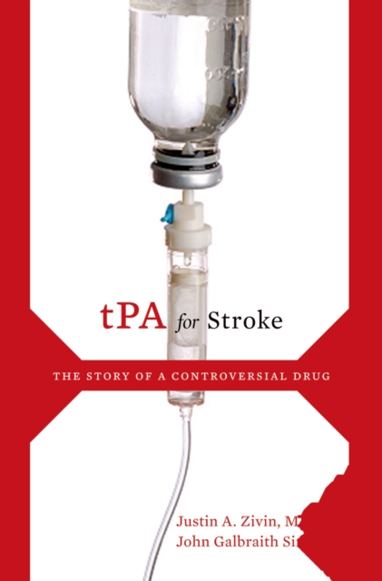
TPA for Stroke
PA for Stroke chronicles how this remarkable drug came to be tested in stroke victims, its early years in development by the pharmaceutical giant Genentech, and its eventual marginalization due to a convergence of unfavorable political, fiscal, and medical circumstances. For instance, initially many stroke specialists were unconvinced that the drug's benefits outweigh its risks (tPA was originally developed and is still used for cardiac patients). Moreover, neurologists called upon to assess stroke patients have not typically been trained to make decisions in emergency settings--and tPA must be given within a scant few hours after stroke. These and other factors have continued to delay the drug's universal acceptance as the most effective treatment available, and to hamper the general public's awareness that such a treatment exists--a troubling state of affairs that Zivin and Simmons argue must be rectified. Instilling the knowledge that anyone, at any time, is susceptible to stroke, from the old and infirm to the young and healthy, tPA for Stroke is a clarion call to awareness in a rapidly changing healthcare environment in which stroke, long a disease in thrall to resignation and pessimism, must be neglected no longer.
eBook
Coagulation Testing: Basic and Advanced Clinical Laboratory Tests
The diagnosis of defects in the hemostatic system leading to bleedings or thromboembolic manifestations is, to a great extent, dependent on laboratory assays capable of identifying the molecular and/or functional defects. A thorough medical as well as drug history is essential in order to perform a relevant laboratory investigation. Is the defect most likely hereditary or acquired? Could the symptoms be drug induced? A good medical history can also give an idea which part of the hemostatic system that could be involved. Laboratory tests are then vital for verification of the suspicion. In this chapter, we give an overview of commonly used screening tests as well as specific analyses.
Book Chapter


DNA liquid biopsy-based prediction of cancer-associated venous thromboembolism
Cancer-associated venous thromboembolism (VTE) is a major source of oncologic cost, morbidity and mortality. Identifying high-risk patients for prophylactic anticoagulation is challenging and adds to clinician burden. Circulating tumor DNA (ctDNA) sequencing assays (‘liquid biopsies’) are widely implemented, but their utility for VTE prognostication is unknown. Here we analyzed three plasma sequencing cohorts: a pan-cancer discovery cohort of 4,141 patients with non-small cell lung cancer (NSCLC) or breast, pancreatic and other cancers; a prospective validation cohort consisting of 1,426 patients with the same cancer types; and an international generalizability cohort of 463 patients with advanced NSCLC. ctDNA detection was associated with VTE independent of clinical and radiographic features. A machine learning model trained on liquid biopsy data outperformed previous risk scores (discovery, validation and generalizability c-indices 0.74, 0.73 and 0.67, respectively, versus 0.57, 0.61 and 0.54 for the Khorana score). In real-world data, anticoagulation was associated with lower VTE rates if ctDNA was detected (
n
= 2,522, adjusted hazard ratio (HR) = 0.50, 95% confidence interval (CI): 0.30–0.81); ctDNA
−
patients (
n
= 1,619) did not benefit from anticoagulation (adjusted HR = 0.89, 95% CI: 0.40–2.0). These results provide preliminary evidence that liquid biopsies may improve VTE risk stratification in addition to clinical parameters. Interventional, randomized prospective studies are needed to confirm the clinical utility of liquid biopsies for guiding anticoagulation in patients with cancer.
An analysis of three multi-cancer cohorts shows that the detection of ctDNA using liquid biopsies has the potential to improve the risk prediction of cancer-associated venous thromboembolism.
Journal Article

Assessing the exercise‐related kinetics of circulating cell‐free DNA, circulating tumour DNA, DNase I activity and cytokines in patients with solid tumours: A pilot study
Circulating cell‐free DNA (cfDNA), circulating tumour DNA (ctDNA) and inflammatory cytokines have prognostic and predictive value in oncology. However, the effects of acute exercise on cfDNA levels are unknown. Here, we explore the kinetics of cfDNA, ctDNA and cytokines upon an incremental exercise test in a pilot cohort of cancer patients compared with healthy control subjects. Patients with solid tumours (n = 12) and age‐matched control subjects (n = 6) were recruited to perform an all‐out cardiopulmonary bicycle test. Blood samples were collected before (Pre), directly after (Post) and 90 min after the test (+90 min), and the cfDNA, ctDNA (Kirsten rat sarcoma viral oncogene homolog (KRAS) mutations), DNase I activity and cytokine levels were measured. Cardiopulmonary exercise testing was easily feasible in cancer patients, and data from eight patients and five control subjects were available for exploratory statistical evaluation. The cfDNA levels increased from Pre to Post and decreased to baseline at +90 min in all subjects. The cfDNA concentrations and DNase I activity were clearly correlated in the control but not in the cancer group. Neutrophil‐associated myeloperoxidase (MPO), calprotectin (MRP 8/14), and lipocalin A (NGAL) showed strong responses to exercise. The percentage of ctDNA, detected in only one cancer patient, decreased after acute exercise. In our study, we could safely perform cardiopulmonary exercise testing with patients with different cancer entities and subsequently run biomarker analyses. Our results hint at an exercise‐triggered release of cfDNA and neutrophil‐derived cytokines in cancer patients. What is the central question of this study? How does acute exercise affect the release and clearance of circulating cell‐free DNA in patients with solid tumours in comparison to healthy control subjects? What is the main finding and its importance? Incremental exercise immediately increases the levels of circulating cell‐free DNA and the DNase I activity in patients and control subjects. Healthy subjects show larger increases and decreases, whereas, importantly, in patients the levels normalize after 90 min of rest. Given that exercise can affect the proportion of circulating tumour DNA, our results highlight the importance of considering exercise as a preanalytical factor for cancer liquid biopsies.
Journal Article

The Impact of the Anticoagulant Type in Blood Collection Tubes on Circulating Extracellular Plasma MicroRNA Profiles Revealed by Small RNA Sequencing
Pre-analytical factors have a significant influence on circulating microRNA (miRNA) profiling. The aim of this study was a comprehensive assessment of the impact of the anticoagulant type in blood collection tubes on circulating plasma miRNA profiles using small RNA sequencing. Blood from ten healthy participants (five males and five females from 25 to 40 years old) was taken in collection tubes with four different anticoagulants: acid citrate dextrose (ACD-B), sodium citrate, citrate-theophylline-adenosine-dipyridamole (CTAD) and dipotassium-ethylenediaminetetraacetic acid (K2 EDTA). Platelet-free plasma samples were obtained by double centrifugation. EDTA plasma samples had elevated levels of hemolysis compared to samples obtained using other anticoagulants. Small RNA was extracted from plasma samples and small RNA sequencing was performed on the Illumina NextSeq 500 system. A total of 30 samples had been successfully sequenced starting from ~1 M reads mapped to miRNAs, allowing us to analyze their diversity and isoform content. The principal component analysis showed that the EDTA samples have distinct circulating plasma miRNA profiles compared to samples obtained using other anticoagulants. We selected 50 miRNA species that were differentially expressed between the sample groups based on the type of anticoagulant. We found that the EDTA samples had elevated levels of miRNAs which are abundant in red blood cells (RBC) and associated with hemolysis, while the levels of some platelet-specific miRNAs in these samples were lowered. The ratio between RBC-derived and platelet-derived miRNAs differed between the EDTA samples and other sample groups, which was validated by quantitative PCR. This study provides full plasma miRNA profiles of 10 healthy adults, compares them with previous studies and shows that the profile of circulating miRNAs in the EDTA plasma samples is altered primarily due to an increased level of hemolysis.
Journal Article