Catalogue Search | MBRL


Are you sure you want to remove the book from the shelf?

{{itemTitle}}
1,663
result(s) for
"Corneal Transplantation - methods"
Sort by:
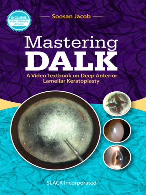
Mastering DALK
Due to its instantaneous and visible results for patients both anatomically and visually, anterior lamellar keratoplasty is a field of great interest to corneal surgeons. Newer techniques of DALK, femtosecond assisted DALK, TILK, and more have inherent advantages that make them supersede older techniques of keratoplasty by far.
Mastering DALK: A Video Textbook on Deep Anterior Lamellar Keratoplasty
by Dr. Soosan Jacob combines the practical explanation of written text with the dynamic demonstration of surgical videos.
From pre-operative to post-operative patient management and every surgical technique in between, this unique book and its included video website is a complete education on DALK. This includes the history and basics of lamellar keratoplasty, as well as tutorials on various surgical techniques currently in use, advice for complex situations, and management of possible complications.
Each chapter includes high-quality photographs and illustrations and is paired with videos on the accompanying website to demonstrate techniques and provide step-by-step instructions. The surgeries in these videos are performed and narrated by the same team of leading international experts who have also written all of the clinical tips, tricks, and pearls of wisdom throughout the book.
Some videos include:
Retrieving the Lost Bubble
DALK Surgery with Type 1 Big Bubble
Groove Peel DALK
Femtosecond Laser Assisted DALK
SMILE Away a Dermoid
Any surgeon looking to add anterior lamellar keratoplasty to their practice will appreciate the digestible nature of
Mastering DALK: A Video Textbook on Deep Anterior Lamellar Keratoplasty,
which provides both the crucial detail of a book and the irreplaceable value of visual demonstration.
eBook

A study protocol for a multicentre randomised clinical trial evaluating the safety and feasibility of a bioengineered human allogeneic nanostructured anterior cornea in patients with advanced corneal trophic ulcers refractory to conventional treatment
IntroductionThere is a need to find alternatives to the use of human donor corneas in transplants because of the limited availability of donor organs, the incidence of graft complications, as well as the inability to successfully perform corneal transplant in patients presenting limbal deficiency, neo-vascularized or thin corneas, etc. We have designed a clinical trial to test a nanostructured fibrin-agarose corneal substitute combining allogeneic cells that mimics the anterior human native cornea in terms of optical, mechanical and biological behaviour.Methods and analysisThis is a phase I-II, randomised, controlled, open-label clinical trial, currently ongoing in ten Spanish hospitals, to evaluate the safety and feasibility, as well as clinical efficacy evidence, of this bioengineered human corneal substitute in adults with severe trophic corneal ulcers refractory to conventional treatment, or with sequelae of previous ulcers. In the initial phase of the trial (n=5), patients were sequentially recruited, with a safety period of 45 days, receiving the bioengineered corneal graft. In the second phase of the trial (currently ongoing), subjects are block randomised (2:1) to receive either the corneal graft (n=10), or amniotic membrane (n=5), as the control treatment. Adverse events, implant status, infection signs and induced neovascularization are evaluated as determinants of safety and feasibility of the bioengineered graft (main outcomes). Study endpoints are measured along a follow-up period of 24 months, including 27 post-implant assessment visits according to a decreasing frequency. Intention to treat, and per protocol, and safety analysis will be performed.Ethics and disseminationThe trial protocol received written approval by the corresponding Ethics Committee and the Spanish Regulatory Authority and is currently recruiting subjects. On completion of the trial, manuscripts with the results of phases I and II of the study will be published in a peer-reviewed journal.Trial registrationCT.gov identifier: NCT01765244 (Jan2013). EudraCT number: 2010-024290-40 (Dec2012).
Journal Article

Comprehensive combined analysis of physician-initiated phase II and III clinical trials on a cultured human corneal endothelial cell product for treating bullous keratopathy
To evaluate the efficacy and safety of a cultured human corneal endothelial cell (cHCEC) product in eyes with bullous keratopathy (BK). Combined analysis of multicenter phase II and III clinical trials. This analysis involved 15 BK eyes in the phase II trial and 12 BK eyes in the phase III trial that underwent cHCEC transplant therapy. Safety was assessed in all the cases. Efficacy was assessed in 17 cases with exclusion of the low- and medium-dose groups in the phase II trial. The primary endpoint was a corneal endothelial cell density of 1000 cells/mm
2
or more at 24 weeks post-transplant, which was attained in 94.1% of the eyes (16 of 17), with a 95% CI of 71.3–99.9%. Additionally, 82.4% of the eyes (14 of 17) met the secondary endpoint of reduction in corneal thickness to less than 630 µm without corneal epithelial edema within the same time frame, with a 95% CI of 56.6–96.2%. The mean decrease in corneal thickness from baseline to 24 weeks post-transplant was −187.4 µm (95% CI, −240.2 µm to −134.5 µm). Furthermore, all the eyes exhibited improvement in best-corrected visual acuity from baseline to 24 weeks post-transplant (95% CI, 80.5–100.0%). By 24 weeks post-transplant, 88.9% of the patients (24 of 27) had experienced adverse events, which were mostly local, mild, and transient. The cHCEC product of this study reconstitutes the corneal endothelial layer with high cellular density and restores corneal thickness and improves visual acuity.
Journal Article

Allogeneic Ex Vivo Expanded Corneal Epithelial Stem Cell Transplantation: A Randomized Controlled Clinical Trial
Limbal stem cell deficiency (LSCD) is a disease resulting from the loss or dysfunction of epithelial stem cells, which seriously impairs sight. Autologous limbal stem cell transplantation is effective in unilateral or partial bilateral disease but not applicable in total bilateral disease. An allogeneic source of transplantable cells for use in total bilateral disease can be obtained from culture of donated cadaveric corneal tissue. We performed a controlled multicenter study to examine the feasibility, safety, and efficacy of allogeneic corneal epithelial stem cells in the treatment of bilateral LSCD. Patients were randomized to receive corneal epithelial stem cells cultured on amniotic membrane (AM): investigational medicinal product (IMP) or control AM only. Patients received systemic immunosuppression. Primary endpoints were safety and visual acuity, secondary endpoint was change in composite ocular surface score (OSS). Sixteen patients were treated and 13 patients completed all assessments. Safety was demonstrated and 9/13 patients had improved visual acuity scores at the end of the trial, with no significant differences between IMP and control groups. Patients in the IMP arm demonstrated significant, sustained improvement in OSS, whereas those in the control arm did not. Serum cytokine levels were measured during and after the period of immune suppression and we identified strongly elevated levels of CXCL8 in the serum of patients with aniridia, which persisted throughout the trial. This first randomized control trial of allogeneic corneal epithelial stem cells in severe bilateral LSCD demonstrates the feasibility and safety of this approach. Stem Cells Translational Medicine 2019;8:323–331 Patients with severe ocular surface disorder received transplants of amniotic membrane with (black bars) or without (gray bars) cadaveric‐donor‐derived cultured limbal stem cells. All patients received immune suppression. Only patients who received transplants containing limbal stem cells showed sustained significant improvements (reductions) in combined ocular surface scores (5 factors scored 0–3 where 0 is a normal eye score).
Journal Article

Large-diameter deep anterior lamellar keratoplasty for keratoconus: visual and refractive outcomes
BackgroundTo investigate the difference in clinical outcomes between large-diameter deep anterior lamellar keratoplasty (L-DALK) and standard DALK (S-DALK) for the treatment of keratoconus.Methods132 patients (132 eyes) from the Zhongshan Ophthalmic Center with a clinical diagnosis of keratoconus were enrolled. The participants were featured by the intolerance to rigid gas-permeable contact lenses or unsuccessful fitting of contact lenses. Using stratified blocked randomisation, eligible eyes were allocated into the L-DALK group or the S-DALK group (66 eyes, respectively). Postoperative uncorrected visual acuity (UCVA), best spectacle-corrected visual acuity (BSCVA), refractive sphere, manifest cylinder and spherical equivalent refractive error were tested at 6, 12, 18 and 24 months after surgery.ResultsAfter surgery, the L-DALK group had better UCVA and BSCVA than the S-DALK group (p=0.000 and 0.021, respectively). At 24 months, mean BSCVA was 0.17±0.10 logarithm of the minimum angle of resolution (logMAR) (Snellen equivalent, 20/25) in the L-DALK group vs 0.22±0.10 logMAR (Snellen equivalent, 20/32) in the S-DALK group. Differences were observed between the L-DALK group and the S-DALK group in terms of refractive sphere (p=0.015), manifest cylinder (p=0.014) and spherical equivalent refractive error (p=0.034) at any time interval postoperatively. At 24 months, the mean spherical equivalent refractive error was −3.5±3.2 D and −5.2±2.6 D in the L-DALK and S-DALK groups, respectively.ConclusionsL-DALK can reduce the degree of postoperative myopia and manifest astigmatism and improve visual acuity outcomes in keratoconus compared with S-DALK.Trial registration numberChinese Clinical Trial Registry (TRC-13003122).
Journal Article

Femtosecond Laser Assisted Deep Anterior Lamellar Keratoplasty Outcomes and Healing Patterns Compared to Manual Technique
The purpose of the study is to report the visual, refractive, and wound healing pattern outcomes of femtosecond assisted deep anterior lamellar keratoplasty (DALK) compared to the conventional manual technique. DALK was performed on 50 eyes of 47 advanced keratoconus patients. The patients were divided into two groups, 25 eyes each, depending on whether femtosecond assisted or manual DALK technique was performed for the side cut of the procedure only. Patients were followed up at 1 month, 6 months, and 1 year for visual acuity, clinical refraction, corneal cylinder, date of suture removal, and side cut corneal healing pattern according to new grading classification of the side cut scar (Grade 0 = transparent scar, 1 = faint healing opacity, 2 = evident healing opacity, 3 = significant opacity with some cosmetic imbalance, and 4 = highly significant opacity with very significant cosmetic imbalance). Outcomes are reported at one year. In conclusion, femtosecond assisted and manual DALK show comparable visual and refractive outcomes but femtosecond assisted DALK shows more evident corneal wound healing patterns at the side cut. This observation may indicate that an activated cornea wound healing might allow earlier suture removal when femtosecond technology is used to perform the side cut for DALK.
Journal Article
Long-term comparison of full-bed deep anterior lamellar keratoplasty and penetrating keratoplasty in treating keratoconus
Objective: To compare postoperative outcomes of full-bed deep anterior lamellar keratoplasty (DALK) with penetrating keratoplasty (PK) in treating keratoconus. Methods: Seventy-five eyes of 64 patients who received full-bed DALK and 52 eyes of 51 patients who received PK between June 2000 and August 2010 were included in this ret- rospective study. Full-bed DALK was performed using Yao's hooking-detaching technique. PK was performed using a standard technique. Intraoperative and postoperative complications, visual acuity, rejection, graft survival, endothelial cell density, corneal sensation recovery, and re-innervation were compared between the two groups. Results: A best correct visual acuity of 0.5 or better was achieved in 90.7% of eyes after full-bed DALK and in 92.3% of eyes after PK (P=0.75). By the fifth postoperative year, graft endothelial cell loss reached 34.6% in the PK group vs. 13.9% in the full-bed DALK group (P〈0.001). There were no statistical differences in corneal sensitivity recovery or corneal re-innervation between the groups (P〉0.05). Intraoperative microperforation occurred in seven out of 75 (9.3%) eyes with a temporally postoperative double anterior chamber in two eyes in the full-bed DALK group. Postoperative com- plications in the PK vs. the full-bed DALK groups respectively were: rejection (7.7% vs. 0%, P=0.015), high intraocular pressure (lOP) (46.2% vs. 1.3%, P〈0.001), secondary glaucoma (9.6% vs. 0%, P=0.006), complicated cataract (19.2% vs. 0%, P〈0.001), and wound dehiscence (9.6% vs. 0%, P=0.006). Conclusions: Both full-bed DALK and PK can offer long-term satisfactory visual outcomes for keratoconus. Graft rejection, secondary glaucoma, complicated cataracts, and constant endothelial cell loss were observed in eyes only after PK.
Journal Article

Subconjunctival and/or intrastromal bevacizumab injections as preconditioning therapy to promote corneal graft survival
The purpose of this study is to investigate whether subconjunctival and/or intrastromal Bevacizumab injections could help to prevent graft failure in high-risk keratoplasties. Twenty seven eyes of 27 patients, affected by high immune rejection risk and corneal neovascularization, were involved in this prospective interventional case–control series (case group: 14 eyes and control group: 13 eyes). Case group was submitted to a cycle of three subconjunctival and/or intrastromal injections of 5 mg/0.2 ml Bevacizumab. After a mean period of 6.36 months ± 3.38 SD from the last injection, all patients underwent keratoplasty. An adjunctive injection was performed intraoperatively at the end of the surgical procedure. Control group did not receive any Bevacizumab injection, but directly underwent keratoplasty. Each patient was submitted to a complete eye examination and corneal confocal microscopy. The absence of immune rejection signs in the graft, at clinical and confocal microscopy examination, was considered as main outcome measure. All cases showed less ocular inflammation and activity of vessels. No side effects were detected after the injection procedure. No corneal graft rejection was seen during the follow-up (mean 26.1 months ± 5.7 SD) in the case group. Six eyes of the control group showed graft rejection 3.8 months ± 1.4 SD after keratoplasty. As a conclusion, Bevacizumab injection may represent a preconditioning treatment to improve prognosis in high-risk corneal transplantation. The procedure seems to be safe and it may help to reduce the inflammatory stimulus that plays a key role in corneal graft rejection.
Journal Article
Descemet’s membrane endothelial keratoplasty is the predominant keratoplasty procedure in Germany since 2016: a report of the DOG-section cornea and its keratoplasty registry
Background/aimsThis retrospective multicentric panel study provides absolute numbers, types of and indications for corneal transplantation in Germany from 2011 to 2021 and sets them into the international context.MethodsA questionnaire was sent to the 104 German ophthalmologic surgery departments and 93 (89%) provided their data.ResultsThe number of reported keratoplasties more than doubled from 2011 (n=4474) to 2021 (n=8998). Lamellar keratoplasties (49% posterior (n=2883), 4% anterior (n=231)) surpassed penetrating keratoplasty (PKP, 47%, n=2721) for the first time in 2014. Since 2016, Descemet’s membrane endothelial keratoplasty (DMEK) has become the predominant keratoplasty procedure in Germany. Its number increased by 1.5-fold from 3850 (2016) to 5812 (2021). Main indications in 2021 were Fuchs’ endothelial corneal dystrophy (FECD, 43%), pseudophakic corneal decompensation (12%), repeated keratoplasty (11%), infections (7%), keratoconus (6%) and corneal scarring (4%, others: 9%). The PKP percentage decreased from 70.2% in 2011 (n=3141) to 31.7% in 2021 (n=2853). Descemet’s stripping (automated) endothelial keratoplasties (DSAEKs) decreased to 1% in 2021 (n=74). 98.6% of all posterior lamellar keratoplasties were DMEKs in Germany in 2021. The number of deep anterior lamellar keratoplasties (DALKs) remained comparable from 2011 (n=269) to 2021 (n=251, 2.8%).ConclusionMain indications for corneal transplantation in Germany (2021) were FECD and pseudophakic corneal decompensation. DMEK is by far the predominant corneal transplantation procedure since 2016 followed by PKP, whose absolute number decreased only slightly during the decade from 2011 to 2021. DALK proportions remain low, but stable, whereas DSAEK decreased annually and plays a minor role in Germany.Trial registration number NCT03381794.
Journal Article

Corneal transplantation
Corneal transplantation or keratoplasty has developed rapidly in the past 10 years. Penetrating keratoplasty, a procedure consisting of full-thickness replacement of the cornea, has been the dominant procedure for more than half a century, and successfully caters to most causes of corneal blindness. The adoption by specialist surgeons of newer forms of lamellar transplantation surgery, which selectively replace only diseased layers of the cornea, has been a fundamental change in recent years. Deep anterior lamellar keratoplasty is replacing penetrating keratoplasty for disorders affecting the corneal stromal layers, while eliminating the risk of endothelial rejection. Endothelial keratoplasty, which selectively replaces the corneal endothelium in patients with endothelial disease, has resulted in more rapid and predictable visual outcomes. Other emerging therapies are ocular surface reconstruction and artificial cornea (keratoprosthesis) surgery, which have become more widely available because of rapid advances in these techniques. Collectively, these advances have resulted in improved outcomes, and have expanded the number of cases of corneal blindness, which can now be treated successfully. Femtosecond-laser-assisted surgery, bioengineered corneas, and medical treatment for endothelial disease are also likely to play a part in the future.
Journal Article