Catalogue Search | MBRL


Are you sure you want to remove the book from the shelf?

{{itemTitle}}
2,635
result(s) for
"Hand Injuries surgery."
Sort by:


A randomized clinical trial comparing early active motion programs: Earlier hand function, TAM, and orthotic satisfaction with a relative motion extension program for zones V and VI extensor tendon repairs
Randomized clinical trial with parallel groups.
Early active mobilization programs are used after zones V and VI extensor tendon repairs; two programs used are relative motion extension (RME) orthosis and controlled active motion (CAM). Although no comparative studies exist, use of the RME orthosis has been reported to support earlier hand function.
This randomized clinical trial investigated whether patients managed with an RME program would recover hand function earlier postoperatively than those managed with a CAM program.
Forty-two participants with zones V-VI extensor tendon repairs were randomized into either a CAM or RME program. The Sollerman Hand Function Test (SHFT) was the primary outcome measure of hand function. Days to return to work, QuickDASH (Disabilities of Arm, Shoulder and Hand) questionnaire, total active motion (TAM), grip strength, and patient satisfaction were the secondary measures of outcome.
The RME group demonstrated better results at four weeks for the SHFT score (P = .0073; 95% CI: −10.9, −1.8), QuickDASH score (P = .05; 95% CI: −0.05, 19.5), and TAM (P = .008; 95% CI: −65.4, −10.6). Days to return to work were similar between groups (P = .77; 95% CI: −28.1, 36.1). RME participants were more satisfied with the orthosis (P < .0001; 95% CI: 3.5, 8.4). No tendon ruptures occurred.
Participants managed using an RME program, and RME finger orthosis demonstrated significantly better early hand function, TAM, and orthosis satisfaction than those managed by the CAM program using a static wrist-hand-finger orthosis. This is likely due to the less restrictive design of the RME orthosis.
The RME program supports safe earlier recovery of hand function and motion when compared to a CAM program following repair of zones V and VI extensor tendons.
•This randomized clinical trial compared relative motion extension to controlled active motion.•A relative motion extension program enabled earlier return to hand function.•Patients were more satisfied with a relative motion extension finger orthosis.•A relative motion extension program did not result in earlier return to work.
Journal Article

Antegrade Intramedullary Pinning Versus Retrograde Intramedullary Pinning for Displaced Fifth Metacarpal Neck Fractures
Background
Severe angulation or shortening can be a surgical indication for fifth metacarpal neck fracture. In a previous meta-analysis, antegrade intramedullary pinning was shown to produce better hand function outcomes than percutaneous transverse pinning or miniplate fixation for treatment of fifth metacarpal neck fractures. However, the outcomes of retrograde intramedullary pinning, to our knowledge, have not been compared with those of antegrade intramedullary pinning.
Questions/purposes
We asked whether the clinical and radiographic outcomes of antegrade intramedullary pinning are different from those of percutaneous retrograde intramedullary pinning for treating patients with displaced fifth metacarpal neck fractures.
Methods
Forty-six patients with displaced fifth metacarpal neck fractures with an apex dorsal angulation greater than 30° were enrolled in our prospective study. Subjects were treated randomly by antegrade intramedullary pinning (antegrade group) or by percutaneous retrograde intramedullary pinning (retrograde group). Clinical evaluations, which included active ROM of the fifth metacarpophalangeal joint, VAS for pain, grip strength, and DASH score, were performed at 3 months and 6 months postoperatively. Radiographic evaluations of apex dorsal angulation and axial shortening were performed preoperatively and 6 months postoperatively.
Results
Patients in the antegrade group achieved better outcomes than patients in the retrograde group for all clinical parameters at 3 months postoperatively (ROM: antegrade median 80° [range, 57°–90°] versus retrograde 69° [range, 45°–90°], difference of medians 11°, p < 0.001; VAS: antegrade median of 2 [range, 0–5] versus retrograde 4 [range, 0–7], difference of medians 2, p < 0.001; grip strength: antegrade median 81% [range, 60%–100%] versus retrograde 71% [range, 49%–98%], differences of medians 10%, p < 0.001; DASH: antegrade median 4.3 [range, 0–15.8] versus retrograde 10.3 [range, 0–28.4], difference of medians 6, p < 0.001), but these differences, with the numbers available, were not observed at 6 months postoperatively for any clinical parameters (ROM: antegrade median 88° [range, 81°–90°] versus retrograde 87° [range, 80°–90°], difference of medians 1°, p = 0.35; VAS: antegrade median 1 [range, 0–2] versus retrograde 1[range, 0–3], difference of medians 0, p = 0.67; grip strength: antegrade median 93% [range, 78%–104%] versus retrograde 91% [range, 76%–101%], difference of medians 2%, p = 0.41; DASH: antegrade median 3 [range, 0–12.5] versus retrograde of 4.3 [range, 0–15.8], difference of medians 1.3, p = 0.48). At 6 months postoperatively, there also were no differences, with the numbers available, in radiographic parameters between the antegrade and retrograde fixation groups. Residual angulation was not different (antegrade median: 7° [range, 2°–11°], retrograde: 9° [range, 3°–13°], difference of medians 2°, p = 0.56). Shortening between the two groups also was not different (antegrade median: 1 mm [range, 0 mm–2 mm], retrograde median: 1 mm [range, 0 mm–2 mm], difference of medians 0, p = 0.78).
Conclusion
Our study findings suggest antegrade intramedullary pinning has some clinical advantages during the early recovery period over percutaneous retrograde intramedullary pinning for treatment of displaced fifth metacarpal neck fractures, but the advantages are not evident at 6 months postoperatively. In addition, our study showed no differences in radiographic outcomes between antegrade and retrograde techniques. For patients who require an early return of hand function, such as athletes, antegrade intramedullary pinning can be recommended. Otherwise, treatment could be decided according to the surgeon’s preference and patient status, and based on consideration of the need for an accessory procedure for pin removal after antegrade intramedullary pinning.
Level of Evidence
Level I, therapeutic study.
Journal Article

Comparison of functional metacarpal splint and ulnar gutter splint in the treatment of fifth metacarpal neck fractures: a prospective comparative study
Background
Fifth metacarpal fractures are the most common fractures of the hand. These fractures are generally treated with conservative methods. The aim of this study was to compare the radiological and clinical outcomes of two conservative treatment methods, functional metacarpal splint(FMS) and ulnar gutter splint(UGS), for the treatment of fifth metacarpal neck fractures.
Methods
A prospective comparative study was designed to assess the conservative treatment of isolated and closed stable fractures of the fifth metacarpal neck. In total, 58 patients were included in the study and were treated with FMS or UGS after fracture reduction in a consecutive order. Angulation, shortening and functional outcome (
Quick
DASH scores and grip strengths) were evaluated at the 2nd and 6th months.
Results
Forty patients returned for follow-up. Twenty-two patients were treated with FMS, and 18 patients were treated with UGS. The average age was 28 years (SD ± 12, range;18–43) in the FMS group and 30 years (SD ± 14, range;18–58) in the UGS group. After reduction, significant correction was achieved in both groups, but the average angulation was lower in the FMS group(16 ± 7) compared with the UGS group (21 ± 8)(
p
= 0.043). However, this better initial reduction in FMS group(16 ± 7) could not be maintained in the 1st month follow-up (21 ± 5) (
p
= 0.009). In the FMS group, the improvement in
Quick
DASH scores between the 2nd and 6th month follow-up was significant (
p
= 0.003) but not in the UGS group(
p
= 0.075). When the expected grip strengths were calculated, the FMS group reached the expected strength values at the 2nd month follow-up, whereas the UGS group still exhibited significantly lower grip strength at the 2nd month follow-up(
p
= 0.008). However, at the end of the 6th month follow-up, both groups exhibited similar reduction,
Quick
DASH and grip strength values.
Conclusions
In stable 5th metacarpal neck fractures, FMS is adequate to prevent loss of reduction and yields faster improvement in clinical scores with earlier gain of normal grip strength compared with UGS. However, in the long term, both FMS and UGS methods yield similar radiological and clinical outcomes. Patient comfort and compliance may be better with FMS due to less joint restriction, and these findings should be considered when deciding the treatment method.
Trial registration
ISRCTN79534571
The date of registration: 01/04/2019
Type of study/level of evidence:
Therapeutic, II.
Journal Article

Clinical Comparison of Adding Sulfate Magnesium and Dexmedetomidine in Axillary Plexus Block for Prolonging the Duration of Sensory and Motor Block: Study Protocol for a Double-blind Randomized Clinical Trial
Background: The purpose of this study was to compare the effect of magnesium sulfate adjunct to dexmedetomidine on increasing the duration of sensory and motor block in axillary block. Materials and methods: This study is a double-blind clinical trial. Ninety-nine patients were included in the study. They were undergoing forearm and hand surgery and were referred to Vali-e-Asr Hospital in Arak. The patients were divided into three groups. The first group received lidocaine (1.5%) and dexmedetomidine (0.5 μg/kg). The second group patients were given lidocaine (1.5%) plus magnesium. In the control group, lidocaine (1.5%) was adjusted to 35 cc with normal saline. The final volume was 35 cc in the three groups. Sensory and motor block and pain were measured and data were analyzed using SPSS v. 20. The final volume was 35 cc in the three groups. Results: The sensory and motor block onset time and the stabilization time of the sensory and motor block in the magnesium sulfate group were lower (p<0/001). Pain in recovery, 2, 4, 6, 12, and 24 hours after surgery was lower in the magnesium sulfate group when compared with the dexmedetomidine group (p<0.001). The lowest dose of opioid was used in the dexmedetomidine group 24 hours after surgery (p<0.001). Conclusion: The results showed that dexmedetomidine decreases pain. Magnesium sulfate increased the sensory and motor block onset time, and the sensory and motor block stabilization time, but dexmedetomidine increases the motor block duration.
Journal Article

Comparing Three Postoperative Treatment Protocols for Extensor Tendon Repair in Zones V and VI of the Hand
OBJECTIVE. This pilot study compared the effectiveness of 3 postoperative rehabilitation protocols for patients with Zones V and VI extensor tendon lacerations. METHOD. Twenty-seven patients were recruited from 3 sites and randomly assigned to 1 of 3 established treatment protocols: immobilization, early passive motion (EPM), and early active motion (EAM). Outcome measures were collected at 3, 6, and 12 wk after treatment and included total active motion (TAM). RESULTS. At the end of Week 12, data on 24 injured digits of 18 patients were available for analysis. When data at Weeks 3, 6, and 12 were compared, patients in all groups showed steady improvement in TAM, but digits under the EAM treatment improved to a greater extent over time (F[2, 46] = 75.6, p < .001). CONCLUSION. Patients with Zones V and VI extensor tendon injuries treated with the EAM protocol recovered range of motion more rapidly.
Journal Article
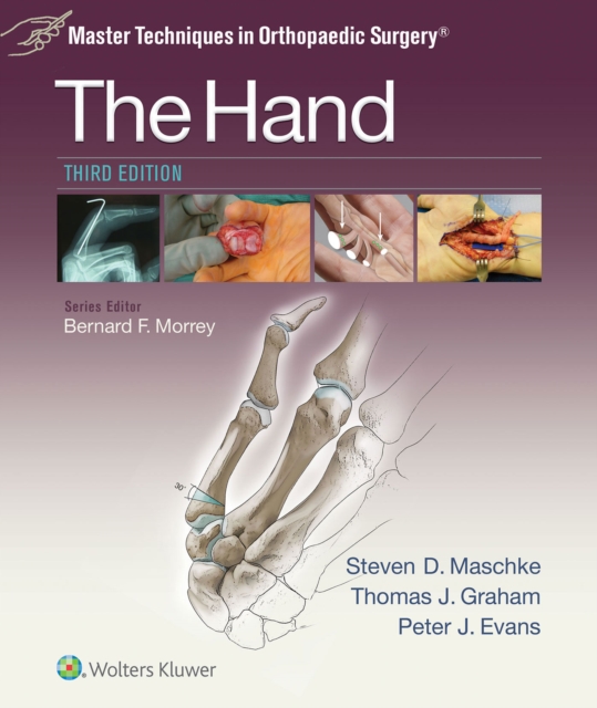
The hand
Master the orthopaedic techniques preferred by today's expert surgeons! The 3rd Edition of this highly regarded title remains your go-to resource for the most advanced and effective surgical techniques for treating traumatic, congenital, inflammatory, neoplastic, and degenerative conditions of the hand. More than 1,000 high-quality photographs and drawings guide you step by step through each procedure, and personal pearls from master surgeons provide operative tips that foster optimal outcomes. 13 new chapters bring you completely up to date with what's new in the field.
eBook

Impact of single versus dual arterial supply on perfusion and function in finger replantation after complex hand injuries
Finger amputations following complex hand injuries (CHI) pose a significant challenge in hand surgery due to severe tissue trauma and neurovascular damage, necessitating precise arterial repair. While restoring arterial perfusion is critical, it remains unclear whether reconstructing both proper palmar digital arteries is required for optimal outcomes. This study evaluates whether restoring one or both arteries in finger replantation after complex injuries impacts perfusion and overall outcomes. In this retrospective, cross-sectional, follow-up study, we investigated patients with finger amputations following CHI admitted to the high-volume regional hand trauma center between January 2013 and December 2020. Perfusion has been assessed using FLIR thermal imaging and laser speckle contrast analysis. Functional outcomes and quality of life scores were measured using standardized questionnaires. Sensory assessments, along with pain and grip strength measurements were also conducted. A total of 31 patients were included in the study. Thermal imaging showed a significantly higher finger surface temperature in patients with two-artery reconstruction. Laser speckle contrast analysis confirmed better perfusion, though not statistically significant. Functional and quality-of-life scores were similar across groups, except for significantly improved temperature sensation in the two-artery group. In conclusion, reconstructing both arteries in finger replantation following CHI isn’t essential for good outcomes if one artery provides adequate perfusion, but dual reconstruction may improve perfusion and temperature sensation.
Journal Article

Sports-related wrist and hand injuries: a review
Background
Hand and wrist injuries are common during athletics and can have a significant impact especially if initially disregarded. Due to their high level of physical demand, athletes represent a unique subset of the population.
Main body
The following is an overview of hand and wrist injuries commonly seen in athletics. Information regarding evaluation, diagnosis, conservative measures, and surgical treatment are provided.
Conclusion
Knowledge of these entities and special consideration for the athlete can help the team physician effectively treat these players and help them achieve their goals.
Journal Article

Diagnosis and treatment of flexor tendon injuries of the hand: what the radiologist needs to know
This article reviews the diagnosis and treatment of flexor tendon injuries of the hand highlighting flexor tendon anatomy, important pre-operative imaging findings, surgical options, and post-operative complications. Imaging plays a key role in guiding treatment of these difficult to manage injuries. Thus, it is important for radiologists to have a sound understanding of factors important in treatment decision-making. In the pre-operative setting, accurately identifying the location of the torn proximal tendon stump in subacute and chronic injuries helps dictate whether the patient is a candidate for a primary flexor tendon repair or may require a tendon reconstruction to restore function. In the post-operative setting, the status of the repair and presence of surrounding adhesions help dictate if and when the patient will require subsequent surgery and whether that surgery will be a tenolysis, revision repair, reconstruction, or fusion.
Journal Article