Catalogue Search | MBRL


Are you sure you want to remove the book from the shelf?

{{itemTitle}}
25,790
result(s) for
"Optical tomography"
Sort by:

OCT or Angiography Guidance for PCI in Complex Bifurcation Lesions
In patients with coronary bifurcation lesions, optical coherence tomography–guided PCI was associated with a lower incidence of major adverse cardiac events at a median 2 years of follow-up than angiography-guided PCI.
Journal Article

Optical coherence tomography-guided versus angiography-guided percutaneous coronary intervention for patients with complex lesions (OCCUPI): an investigator-initiated, multicentre, randomised, open-label, superiority trial in South Korea
Despite the detailed imaging information provided by optical coherence tomography (OCT) during percutaneous coronary intervention (PCI), clinical benefits of this imaging technique in this setting remain uncertain. The aim of the OCCUPI trial was to compare the clinical benefits of OCT-guided versus angiography-guided PCI for complex lesions, assessed as the rate of major adverse cardiac events at 1 year.
This investigator-initiated, multicentre, randomised, open-label, superiority trial conducted at 20 hospitals in South Korea enrolled patients aged 19–85 years for whom PCI with drug-eluting stents was clinically indicated. After diagnostic angiography, clinical and angiographic findings were assessed to identify patients who met the criterion of having one or more complex lesions. Patients were randomly assigned 1:1 to receive PCI with OCT guidance (OCT-guidance group) or angiography guidance without OCT (angiography-guidance group). Web-response permuted-block randomisation (mixed blocks of four or six) was used at each participating site to allocate patients. The allocation sequence was computer-generated by an external programmer who was not involved in the rest of the trial. Outcome assessors were masked to group assignment. Patients, follow-up health-care providers, and data analysers were not masked. PCI was done according to conventional standard methods with everolimus-eluting stents. The primary endpoint was major adverse cardiac events (a composite of cardiac death, myocardial infarction, stent thrombosis, or ischaemia-driven target-vessel revascularisation), 1 year after PCI. The primary analysis was done in the intention-to-treat population. The margin used to establish superiority was 1·0 as a hazard ratio. This trial is registered with ClinicalTrials.gov (NCT03625908) and is completed.
Between Jan 9, 2019, and Sept 22, 2022, 1604 patients requiring PCI with drug-eluting stents for complex lesions were randomly assigned to receive either OCT-guided PCI (n=803) or angiography-guided PCI (n=801). 1290 (80%) of 1604 patients were male and 314 (20%) were female. The median age of patients at randomisation was 64 years (IQR 57–70). 1588 (99%) patients completed 1-year follow-up. The primary endpoint occurred in 37 (5%) of 803 patients in the OCT-guided PCI group and 59 (7%) of 801 patients in the angiography-guided PCI group (absolute difference –2·8% [95% CI –5·1 to –0·4]; hazard ratio 0·62 [95% CI 0·41 to 0·93]; p=0·023). Rates of stroke, bleeding events, and contrast-induced nephropathy were not significantly different across the two groups.
Among patients who required drug-eluting stent implantation for complex lesions, OCT guidance resulted in a lower incidence of major adverse cardiac events at 1 year compared with angiography guidance. These findings indicate the existence of a therapeutic benefit of OCT as an intravascular imaging technique for PCI guidance in patients with complex coronary lesions.
Abbott Vascular and Cardiovascular Research Center.
For the Korean translation of the abstract see Supplementary Materials section.
Journal Article

Line-Field Confocal Optical Coherence Tomography: A New Tool for the Differentiation between Nevi and Melanomas?
Until now, the clinical differentiation between a nevus and a melanoma is still challenging in some cases. Line-field confocal optical coherence tomography (LC-OCT) is a new tool with the aim to change that. The aim of the study was to evaluate LC-OCT for the discrimination between nevi and melanomas. A total of 84 melanocytic lesions were examined with LC-OCT and 36 were also imaged with RCM. The observers recorded the diagnoses, and the presence or absence of the 18 most common imaging parameters for melanocytic lesions, nevi, and melanomas in the LC-OCT images. Their confidence in diagnosis and the image quality of LC-OCT and RCM were evaluated. The most useful criteria, the sensitivity and specificity of LC-OCT vs. RCM vs. histology, to differentiate a (dysplastic) nevus from a melanoma were analyzed. Good image quality correlated with better diagnostic performance (Spearman correlation: 0.4). LC-OCT had a 93% sensitivity and 100% specificity compared to RCM (93% sensitivity, 95% specificity) for diagnosing a melanoma (vs. all types of nevi). No difference in performance between RCM and LC-OCT was observed (McNemar’s p value = 1). Both devices falsely diagnosed dysplastic nevi as non-dysplastic (43% sensitivity for dysplastic nevus diagnosis). The most significant criteria for diagnosing a melanoma with LC-OCT were irregular honeycombed patterns (92% occurrence rate; 31.7 odds ratio (OR)), the presence of pagetoid spread (89% occurrence rate; 23.6 OR) and the absence of dermal nests (23% occurrence rate, 0.02 OR). In conclusion LC-OCT is useful for the discrimination between melanomas and nevi.
Journal Article
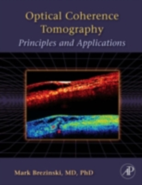
Optical coherence tomography : principles and applications
Optical Coherence Tomography gives a broad treatment of the subject which will include 1)the optics, science, and physics needed to understand the technology 2) a description of applications with a critical look at how the technology will successfully address actual clinical need, and 3) a discussion of delivery of OCT to the patient, FDA approval.
eBook
Optical Coherence Tomography–Guided versus Angiography-Guided PCI
In a randomized trial, optical coherence tomography–guided PCI resulted in a larger minimum stent area than angiography-guided PCI, but there was no between-group difference in target-vessel failure at 2 years.
Journal Article

A feature agnostic approach for glaucoma detection in OCT volumes
Optical coherence tomography (OCT) based measurements of retinal layer thickness, such as the retinal nerve fibre layer (RNFL) and the ganglion cell with inner plexiform layer (GCIPL) are commonly employed for the diagnosis and monitoring of glaucoma. Previously, machine learning techniques have relied on segmentation-based imaging features such as the peripapillary RNFL thickness and the cup-to-disc ratio. Here, we propose a deep learning technique that classifies eyes as healthy or glaucomatous directly from raw, unsegmented OCT volumes of the optic nerve head (ONH) using a 3D Convolutional Neural Network (CNN). We compared the accuracy of this technique with various feature-based machine learning algorithms and demonstrated the superiority of the proposed deep learning based method. Logistic regression was found to be the best performing classical machine learning technique with an AUC of 0.89. In direct comparison, the deep learning approach achieved a substantially higher AUC of 0.94 with the additional advantage of providing insight into which regions of an OCT volume are important for glaucoma detection. Computing Class Activation Maps (CAM), we found that the CNN identified neuroretinal rim and optic disc cupping as well as the lamina cribrosa (LC) and its surrounding areas as the regions significantly associated with the glaucoma classification. These regions anatomically correspond to the well established and commonly used clinical markers for glaucoma diagnosis such as increased cup volume, cup diameter, and neuroretinal rim thinning at the superior and inferior segments.
Journal Article
Choroidal Vascularity Index (CVI) - A Novel Optical Coherence Tomography Parameter for Monitoring Patients with Panuveitis?
To compute choroidal vascularity index (CVI) using an image binarization tool on enhanced depth imaging (EDI)-optical coherence tomography (OCT) scans as a non-invasive optical tool to monitor progression in panuveitis and to investigate the utility of volumetric data from EDI-OCT scans using custom image analysis software.
In this retrospective cohort study, segmented EDI-OCT scans of both eyes in 19 patients with panuveitis were taken at baseline and at 3-month follow-up and were compared with EDI-OCT scans of normal eyes. Subfoveal choroidal area was segmented into luminal (LA) and stromal interstitial area (SA). Choroidal vascularity index (CVI) was defined as the proportion of LA to the total circumscribed subfoveal choroidal area (TCA).
The mean choroidal thickness was 265.5±100.1μm at baseline and 278.4±102.6μm at 3 months follow up (p = 0.06). There was no statistically significant difference in TCA between study and control eyes (p = 0.08). CVI in the control group was 66.9±1.5% at baseline and 66.4±1.5% at follow up. CVI was 74.1±4.7% at baseline and 69.4±4.8% at 3 months follow up for uveitic eyes (p<0.001). The % change in CVI was 6.2 ±3.8 (4.3 to 8.0) for uveitic eyes, which was significantly higher from % change in CVI for control eyes (0.7±1.1, 0.2 to 1.3, p<0.001).
The study reports composite OCT-derived parameters and CVI as a possible novel tool in monitoring progression in panuveitis. CVI may be further validated in larger studies as a novel optical tool to quantify choroidal vascular status.
Journal Article

Functional imaging of the developing brain with wearable high-density diffuse optical tomography: A new benchmark for infant neuroimaging outside the scanner environment
•First demonstration of a wearable, HD-DOT technology, in the infant population.•Advances in system and head-gear design, ergonomics, registration approaches.•Technology is very well tolerated by infant participants.•Production of 3D images of brain function outside the scanner environment.•HD-DOT yields dramatic improvements in spatial specificity and SNR relative to fNIRS.
Studies of cortical function in the awake infant are extremely challenging to undertake with traditional neuroimaging approaches. Partly in response to this challenge, functional near-infrared spectroscopy (fNIRS) has become increasingly common in developmental neuroscience, but has significant limitations including resolution, spatial specificity and ergonomics. In adults, high-density arrays of near-infrared sources and detectors have recently been shown to yield dramatic improvements in spatial resolution and specificity when compared to typical fNIRS approaches. However, most existing fNIRS devices only permit the acquisition of ~20–100 sparsely distributed fNIRS channels, and increasing the number of optodes presents significant mechanical challenges, particularly for infant applications. A new generation of wearable, modular, high-density diffuse optical tomography (HD-DOT) technologies has recently emerged that overcomes many of the limitations of traditional, fibre-based and low-density fNIRS measurements. Driven by the development of this new technology, we have undertaken the first study of the infant brain using wearable HD-DOT. Using a well-established social stimulus paradigm, and combining this new imaging technology with advances in cap design and spatial registration, we show that it is now possible to obtain high-quality, functional images of the infant brain with minimal constraints on either the environment or on the infant participants. Our results are consistent with prior low-density fNIRS measures based on similar paradigms, but demonstrate superior spatial localization, improved depth specificity, higher SNR and a dramatic improvement in the consistency of the responses across participants. Our data retention rates also demonstrate that this new generation of wearable technology is well tolerated by the infant population.
Journal Article
Quantity and quality of image artifacts in optical coherence tomography angiography
To analyze quality and frequency of OCTA artifacts and to evaluate their impact on the interpretability of OCTA images.
75 patients with diabetic retinopathy (DR), retinal artery occlusion (RAO), retinal vein occlusion (RVO), or neovascular age-related macular degeneration (nAMD) and healthy controls were enrolled in this cross-sectional study in the outpatient department of a tertiary eye care center.
All participants underwent an OCTA examination (spectral domain OCT Cirrus 5000 equipped with the AngioPlex module). OCTA scans were analyzed independently by two experienced ophthalmologists. Frequency of various artifacts for the entire OCTA scan and for different segmentation layers and the grading of OCTA interpretability were investigated.
The analysis of 75 eyes of 38 women and 37 men between 24 and 94 years were included. Six eyes had no retinal disease, 19 eyes had nAMD, 16 had DR, 19 eyes had RVO, and 15 eyes showed RAO. A macular edema (ME) was present in 40 of the diseased eyes. Projection artifacts occurred in all eyes in any structure below the superficial retinal vessel layer, segmentation and motion artifacts were found in 55% (41/75) and 49% (37/75) of eyes, respectively. Other artifacts occurred less frequently. Segmentation artifacts were significantly more frequent in diseased than in healthy eyes (p<0.01). Qualitative assessment of OCTA images was graded as excellent in 65% and sufficient in 25% of cases, adding up to 91% images deemed acceptable for examination. Presence of ME was associated with a significantly poorer interpretability (p<0.01).
Various artifacts appear at different frequencies in OCTA images. Nevertheless, a qualitative assessment of the OCTA images is almost always possible. Good knowledge of possible artifacts and critical analysis of the complete OCTA dataset are essential for correct clinical interpretation and determining a precise clinical diagnosis.
Journal Article
Comparison of methods to quantify macular and peripapillary vessel density in optical coherence tomography angiography
To compare macular and peripapillary vessel density values calculated on optical coherence tomography angiography (OCT-A) images with different algorithms, elaborate conversion formula, and compare the ability to discriminate healthy from affected eyes.
Cross-sectional study of healthy subjects, patients with diabetic retinopathy, and glaucoma patients (44 eyes in each group). Vessel density in the macular superficial capillary plexus (SCP), deep capillary plexus (DCP), and the peripapillary radial capillary plexus (RCP) were calculated with seven previously published algorithms. Systemic differences, diagnostic properties, reliability, and agreement of the methods were investigated.
Healthy eyes exhibited higher vessel density values in all plexuses compared to diseased eyes regardless of the algorithm used (p<0.01). The estimated vessel densities were significantly different at all the plexuses (p<0.0001) as a function of method used. Inter-method reliability and agreement was mostly poor to moderate. A conversion formula was available for every method, except for the conversion between multilevel and fixed at the DCP. Substantial systemic, non-constant biases were evident between many algorithms. No algorithm outperformed the others for discrimination of patients from healthy subjects in all the retinal plexuses, but the best performing algorithm varied with the selected plexus.
Absolute vessel density values calculated with different algorithms are not directly interchangeable. Differences between healthy and affected eyes could be appreciated with all methods with different discriminatory abilities as a function of the plexus analyzed. Longitudinal monitoring of vessel density should be performed with the same algorithm. Studies adopting vessel density as an outcome measure should not rely on external normative databases.
Journal Article