Catalogue Search | MBRL


Are you sure you want to remove the book from the shelf?

{{itemTitle}}
1,945
result(s) for
"Pyrophosphates"
Sort by:

Gout and pseudo-gout-related crystals promote GLUT1-mediated glycolysis that governs NLRP3 and interleukin-1β activation on macrophages
ObjectiveMacrophage activation by monosodium urate (MSU) and calcium pyrophosphate (CPP) crystals mediates an interleukin (IL)-1β-dependent inflammation during gout and pseudo-gout flare, respectively. Since metabolic reprogramming of macrophages goes along with inflammatory responses dependently on stimuli and tissue environment, we aimed to decipher the role of glycolysis and oxidative phosphorylation in the IL-1β-induced microcrystal response.MethodsBriefly, an in vitro study (metabolomics and real-time extracellular flux analysis) on MSU and CPP crystal-stimulated macrophages was performed to demonstrate the metabolic phenotype of macrophages. Then, the role of aerobic glycolysis in IL-1β production was evaluated, as well in vitro as in vivo using 18F-fluorodeoxyglucose positron emission tomography imaging and glucose uptake assay, and molecular approach of glucose transporter 1 (GLUT1) inhibition.ResultsWe observed that MSU and CPP crystals led to a metabolic rewiring toward the aerobic glycolysis pathway explained by an increase in GLUT1 plasma membrane expression and glucose uptake on macrophages. Also, neutrophils isolated from human synovial fluid during gout flare expressed GLUT1 at their plasma membrane more frequently than neutrophils isolated from bloodstream. Both glucose deprivation and treatment with either 2-deoxyglucose or GLUT1 inhibitor suppressed crystal-induced NLRP3 activation and IL-1β production, and microcrystal inflammation in vivo.ConclusionIn conclusion, we demonstrated that GLUT1-mediated glucose uptake is instrumental during the inflammatory IL-1β response induced by MSU and CPP crystals. These findings open new therapeutic paths to modulate crystal-related inflammation.
Journal Article

Two bifunctional inositol pyrophosphate kinases/phosphatases control plant phosphate homeostasis
Many eukaryotic proteins regulating phosphate (Pi) homeostasis contain SPX domains that are receptors for inositol pyrophosphates (PP-InsP), suggesting that PP-InsPs may regulate Pi homeostasis. Here we report that deletion of two diphosphoinositol pentakisphosphate kinases VIH1/2 impairs plant growth and leads to constitutive Pi starvation responses. Deletion of phosphate starvation response transcription factors partially rescues vih1 vih2 mutant phenotypes, placing diphosphoinositol pentakisphosphate kinases in plant Pi signal transduction cascades. VIH1/2 are bifunctional enzymes able to generate and break-down PP-InsPs. Mutations in the kinase active site lead to increased Pi levels and constitutive Pi starvation responses. ATP levels change significantly in different Pi growth conditions. ATP-Mg2+ concentrations shift the relative kinase and phosphatase activities of diphosphoinositol pentakisphosphate kinases in vitro. Pi inhibits the phosphatase activity of the enzyme. Thus, VIH1 and VIH2 relay changes in cellular ATP and Pi concentrations to changes in PP-InsP levels, allowing plants to maintain sufficient Pi levels.
Journal Article

Identity and functions of inorganic and inositol polyphosphates in plants
Inorganic polyphosphates (polyPs) and inositol pyrophosphates (PP-InsPs) form important stores of inorganic phosphate and can act as energy metabolites and signaling molecules. Here we review our current understanding of polyP and inositol phosphate (InsP) metabolism and physiology in plants. We outline methods for polyP and InsP detection, discuss the known plant enzymes involved in their synthesis and breakdown, and summarize the potential physiological and signaling functions for these enigmatic molecules in plants.
Journal Article

Cytotoxicity of crystals involves RIPK3-MLKL-mediated necroptosis
Crystals cause injury in numerous disorders, and induce inflammation via the NLRP3 inflammasome, however, it remains unclear how crystals induce cell death. Here we report that crystals of calcium oxalate, monosodium urate, calcium pyrophosphate dihydrate and cystine trigger caspase-independent cell death in five different cell types, which is blocked by necrostatin-1. RNA interference for receptor-interacting protein kinase 3 (RIPK3) or mixed lineage kinase domain like (MLKL), two core proteins of the necroptosis pathway, blocks crystal cytotoxicity. Consistent with this, deficiency of RIPK3 or MLKL prevents oxalate crystal-induced acute kidney injury. The related tissue inflammation drives TNF-α-related necroptosis. Also in human oxalate crystal-related acute kidney injury, dying tubular cells stain positive for phosphorylated MLKL. Furthermore, necrostatin-1 and necrosulfonamide, an inhibitor for human MLKL suppress crystal-induced cell death in human renal progenitor cells. Together, TNF-α/TNFR1, RIPK1, RIPK3 and MLKL are molecular targets to limit crystal-induced cytotoxicity, tissue injury and organ failure.
Kidney stone disease is caused by accumulation of oxalate crystals, which trigger tissue injury, inflammation and cell death. Mulay
et al
. show that crystals induce cell death in the kidney through necroptosis, and propose that this pathway may be a target for the treatment of crystal-induced disease.
Journal Article

Vip1 is a kinase and pyrophosphatase switch that regulates inositol diphosphate signaling
Inositol diphosphates (PP-IPs), also known as inositol pyrophosphates, are high-energy cellular signaling codes involved in nutrient and regulatory responses. We report that the evolutionarily conserved gene product, Vip1, possesses autonomous kinase and pyrophosphatase domains capable of synthesis and destruction of D-1 PP-IPs. Our studies provide atomic-resolution structures of the PP-IP products and unequivocally define that the Vip1 gene product is a highly selective 1-kinase and 1-pyrophosphatase enzyme whose activities arise through distinct active sites. Kinetic analyses of kinase and pyrophosphatase parameters are consistent with Vip1 evolving to modulate levels of 1-IP7 and 1,5-IP8. Individual perturbations in kinase and pyrophosphatase activities in cells result in differential effects on vacuolar morphology and osmotic responses. Analogous to the dual-functional key energy metabolism regulator, phosphofructokinase 2, Vip1 is a kinase and pyrophosphatase switch whose 1-PP-IP products play an important role in a cellular adaptation.
Journal Article
Inositol pyrophosphate dynamics reveals control of the yeast phosphate starvation program through 1,5-IP8 and the SPX domain of Pho81
Eukaryotic cells control inorganic phosphate to balance its role as essential macronutrient with its negative bioenergetic impact on reactions liberating phosphate. Phosphate homeostasis depends on the conserved INPHORS signaling pathway that utilizes inositol pyrophosphates and SPX receptor domains. Since cells synthesize various inositol pyrophosphates and SPX domains bind them promiscuously, it is unclear whether a specific inositol pyrophosphate regulates SPX domains in vivo, or whether multiple inositol pyrophosphates act as a pool. In contrast to previous models, which postulated that phosphate starvation is signaled by increased production of the inositol pyrophosphate 1-IP 7 , we now show that the levels of all detectable inositol pyrophosphates of yeast, 1-IP 7 , 5-IP 7 , and 1,5-IP 8 , strongly decline upon phosphate starvation. Among these, specifically the decline of 1,5-IP 8 triggers the transcriptional phosphate starvation response, the PHO pathway. 1,5-IP 8 inactivates the cyclin-dependent kinase inhibitor Pho81 through its SPX domain. This stimulates the cyclin-dependent kinase Pho85-Pho80 to phosphorylate the transcription factor Pho4 and repress the PHO pathway. Combining our results with observations from other systems, we propose a unified model where 1,5-IP 8 signals cytosolic phosphate abundance to SPX proteins in fungi, plants, and mammals. Its absence triggers starvation responses.
Journal Article

Structural basis for gene regulation by a thiamine pyrophosphate-sensing riboswitch
Riboswitches as drug targets
Genes are commonly turned on or off by protein factors that respond to cellular signals. The recent discovery of riboswitches, regulatory elements within some messenger RNAs, proved that RNA can also detect essential metabolites and control genes. Two structural studies throw new light on the riboswitch system. Serganov
et al
. use X-ray diffraction to establish the three-dimensional structure of a riboswitch from
Escherichia coli
bound to its target, a vitamin B1 derivative. These findings reveal how RNA folds to form a precise pocket for its target and how the antibiotic pyrithiamine acts by tricking the riboswitch. This suggests a new drug design strategy for antibacterials and antifungals targeting riboswitches. Montange and Batey have solved the structure of a bacterial riboswitch RNA bound to
S
-adenosyl methionine. Its complex folded structure reveals how ligand binding leads structural changes that prevent further transcription.
Riboswitches are metabolite-sensing RNAs, typically located in the non-coding portions of messenger RNAs, that control the synthesis of metabolite-related proteins
1
,
2
,
3
. Here we describe a 2.05 Å crystal structure of a riboswitch domain from the
Escherichia coli
thiM
mRNA
4
that responds to the coenzyme thiamine pyrophosphate (TPP). TPP is an active form of vitamin B
1
, an essential participant in many protein-catalysed reactions
5
. Organisms from all three domains of life
6
,
7
,
8
,
9
, including bacteria, plants and fungi, use TPP-sensing riboswitches to control genes responsible for importing or synthesizing thiamine and its phosphorylated derivatives, making this riboswitch class the most widely distributed member of the metabolite-sensing RNA regulatory system. The structure reveals a complex folded RNA in which one subdomain forms an intercalation pocket for the 4-amino-5-hydroxymethyl-2-methylpyrimidine moiety of TPP, whereas another subdomain forms a wider pocket that uses bivalent metal ions and water molecules to make bridging contacts to the pyrophosphate moiety of the ligand. The two pockets are positioned to function as a molecular measuring device that recognizes TPP in an extended conformation. The central thiazole moiety is not recognized by the RNA, which explains why the antimicrobial compound pyrithiamine pyrophosphate targets this riboswitch and downregulates the expression of thiamine metabolic genes. Both the natural ligand and its drug-like analogue stabilize secondary and tertiary structure elements that are harnessed by the riboswitch to modulate the synthesis of the proteins coded by the mRNA. In addition, this structure provides insight into how folded RNAs can form precision binding pockets that rival those formed by protein genetic factors.
Journal Article

Evaluation of Pyrophosphate-Driven Proton Pumps in ISaccharomyces cerevisiae/I under Stress Conditions
In Saccharomyces cerevisiae, pH homeostasis is reliant on ATP due to the use of proton-translocating ATPase (H[sup.+] -ATPase) which constitutes a major drain within cellular ATP supply. Here, an exogenous proton-translocating pyrophosphatase (H[sup.+] -PPase) from Arabidopsis thaliana, which uses inorganic pyrophosphate (PP[sub.i] ) rather than ATP, was evaluated for its effect on reducing the ATP burden. The H[sup.+] -Ppase was localized to the vacuolar membrane or to the cell membrane, and their impact was studied under acetate stress at a low pH. Biosensors (pHluorin and mQueen-2m) were used to observe changes in intracellular pH (pH[sub.i] ) and ATP levels during growth on either glucose or xylose. A significant improvement of 35% in the growth rate at a pH of 3.7 and 6 g·L[sup.−1] acetic acid stress was observed in the vacuolar membrane H[sup.+] -PPase strain compared to the parent strain. ATP levels were elevated in the same strain during anaerobic glucose and xylose fermentations. During anaerobic xylose fermentations, co-expression of pHluorin and a vacuolar membrane H[sup.+] -PPase improved the growth characteristics by means of an improved growth rate (11.4%) and elongated logarithmic growth duration. Our study identified a potential method for improving productivity in the use of S. cerevisiae as a cell factory under the harsh conditions present in industry.
Journal Article
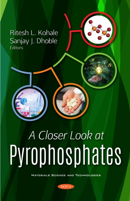
A Closer Look at Pyrophosphates
The present book encompasses and gives brief information about pyrophosphate materials. It also explains their phenomenological theories and properties of rare-earth activated pyrophosphates. This book provides the comprehensive in depth information about the development of phosphates and pyrophosphates along with their essential characterization techniques as well as their diversified applications in numerous domains. This book envelopes all essential information about single and mixed cations pyrophosphate compounds that have been used in past few years. It also covers intact information about innovation in advance pyrophosphate materials especially for their applications in medicinal field. In particular, this book focuses on enhanced chemical and physical properties of pyrophosphate as there is a need for new low-cost, less toxic and energy efficient materials to reduce the current cost of device technologies. This book masterfully demonstrates the present scenario of pyrophosphates and concurrent technologies with its increasing significance to resolve energy- and environment-related problems and their sustainable approach in the world of globalization.
eBook

Emerging roles of inositol pyrophosphates in signaling plant phosphorus status and phytohormone signaling
Phosphorus (P) is an indispensable macronutrient serving a variety of functions in plants. Inositol pyrophosphates (PP-InsPs) nutrient messengers play vital roles in the signaling of P status and plant growth and development. In this review, we summarize (1) the biosynthetic pathway of PP-InsPs and their regulation by plant P status, (2) the effects of PP-InsPs on the function of the SPX domain-containing proteins in signaling plant P status, (3) the effects of inositol pyrophosphates on auxin signaling through TIR1 and on jasmonate signaling through COI1, and (4) the potential crosstalk between P status signaling and phytohormone signaling in plants mediated by inositol pyrophosphates. It is concluded that the interactions between inositol pyrophosphates and their binding proteins are central to plant P status and developmental responses to different P supply.
Journal Article