Catalogue Search | MBRL


Are you sure you want to remove the book from the shelf?

{{itemTitle}}
2,876
result(s) for
"Upper Extremity anatomy "
Sort by:

The Upper Limb of Australopithecus sediba
The evolution of the human upper limb involved a change in function from its use for both locomotion and prehension (as in apes) to a predominantly prehensile and manipulative role. Well-preserved forelimb remains of 1.98-million-year-old Australopithecus sediba from Malapa, South Africa, contribute to our understanding of this evolutionary transition. Whereas other aspects of their postcranial anatomy evince mosaic combinations of primitive (australopith-like) and derived ( Homo -like) features, the upper limbs (excluding the hand and wrist) of the Malapa hominins are predominantly primitive and suggest the retention of substantial climbing and suspensory ability. The use of the forelimb primarily for prehension and manipulation appears to arise later, likely with the emergence of Homo erectus .
Journal Article

Standardizing nomenclature in regional anesthesia: an ASRA-ESRA Delphi consensus study of upper and lower limb nerve blocks
BackgroundInconsistent nomenclature and anatomical descriptions of regional anesthetic techniques hinder scientific communication and engender confusion; this in turn has implications for research, education and clinical implementation of regional anesthesia. Having produced standardized nomenclature for abdominal wall, paraspinal and chest wall regional anesthetic techniques, we aimed to similarly do so for upper and lower limb peripheral nerve blocks.MethodsWe performed a three-round Delphi international consensus study to generate standardized names and anatomical descriptions of upper and lower limb regional anesthetic techniques. A long list of names and anatomical description of blocks of upper and lower extremities was produced by the members of the steering committee. Subsequently, two rounds of anonymized voting and commenting were followed by a third virtual round table to secure consensus for items that remained outstanding after the first and second rounds. As with previous methodology, strong consensus was defined as ≥75% agreement and weak consensus as 50%–74% agreement.ResultsA total of 94, 91 and 65 collaborators participated in the first, second and third rounds, respectively. We achieved strong consensus for 38 names and 33 anatomical descriptions, and weak consensus for five anatomical descriptions. We agreed on a template for naming peripheral nerve blocks based on the name of the nerve and the anatomical location of the blockade and identified several areas for future research.ConclusionsWe achieved consensus on nomenclature and anatomical descriptions of regional anesthetic techniques for upper and lower limb nerve blocks, and recommend using this framework in clinical and academic practice. This should improve research, teaching and learning of regional anesthesia to eventually improve patient care.
Journal Article
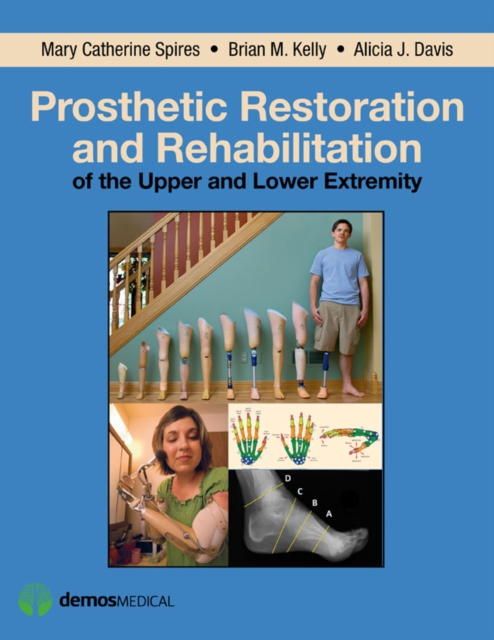
Prosthetic restoration and rehabilitation of the upper and lower extremity
Prosthetic Restoration and Rehabilitation of the Upper and Lower Extremity is a well-illustrated, state-of-the-art reference on the science and practice of post-amputation care, prosthetic restoration, and functional rehabilitation, designed to maximize patient independence and quality of life.
eBook

Investigating the impact of captivity and domestication on limb bone cortical morphology: an experimental approach using a wild boar model
The lack of bone morphological markers associated with the human control of wild animals has prevented the documentation of incipient animal domestication in archaeology. Here, we assess whether direct environmental changes (i.e. mobility reduction) could immediately affect ontogenetic changes in long bone structure, providing a skeletal marker of early domestication. We relied on a wild boar experimental model, analysing 24 wild-born specimens raised in captivity from 6 months to 2 years old. The shaft cortical thickness of their humerus was measured using a 3D morphometric mapping approach and compared with 23 free-ranging wild boars and 22 pigs from different breeds, taking into account sex, mass and muscle force differences. In wild boars we found that captivity induced an increase in cortical bone volume and muscle force, and a topographic change of cortical thickness associated with muscular expression along a phenotypic trajectory that differed from the divergence induced by selective breeding. These results provide an experimental proof of concept that changes in locomotor behaviour and selective breeding might be inferred from long bones morphology in the fossil and archaeological record. These trends need to be explored in the archaeological record and further studies are required to explore the developmental changes behind these plastic responses. Exploring the process of domestication as an integration of animals into human society provides a unique insight into one of the key steps in Homo sapiens evolution, at the root of its global impact over the biosphere 1 and species evolution 2. However, documenting this process, as an intensification of the relationship between humans and animals in archaeology 3 , is challenging 4,5. One of the main issues is that no relevant methodological approach has been able to capture this elusive process. Bioarchaeologists have relied on a morphological 'syndrome of OPEN 1 Archéozoologie, Archéobotanique: Sociétés, Pratiques et Environnements, UMR 7209,
Journal Article

The Tubercles on Humpback Whales' Flippers: Application of Bio-Inspired Technology
Synopsis The humpback whale (Megaptera novaeangliae) is exceptional among the large baleen whales in its ability to undertake aquabatic maneuvers to catch prey. Humpback whales utilize extremely mobile, wing-like flippers for banking and turning. Large rounded tubercles along the leading edge of the flipper are morphological structures that are unique in nature. The tubercles on the leading edge act as passive-flow control devices that improve performance and maneuverability of the flipper. Experimental analysis of finite wing models has demonstrated that the presence of tubercles produces a delay in the angle of attack until stall, thereby increasing maximum lift and decreasing drag. Possible fluid-dynamic mechanisms for improved performance include delay of stall through generation of a vortex and modification of the boundary layer, and increase in effective span by reduction of both spanwise flow and strength of the tip vortex. The tubercles provide a bio-inspired design that has commercial viability for wing-like structures. Control of passive flow has the advantages of eliminating complex, costly, high-maintenance, and heavy control mechanisms, while improving performance for lifting bodies in air and water. The tubercles on the leading edge can be applied to the design of watercraft, aircraft, ventilation fans, and windmills.
Journal Article

Patterns of morphological integration in the appendicular skeleton of mammalian carnivores
We investigated patterns of evolutionary integration in the appendicular skeleton of mammalian carnivores. The findings are discussed in relation to performance selection in terms of organismal function as a potential mechanism underlying integration. Interspecific shape covariation was quantified by two‐block partial least‐squares (2B‐PLS) analysis of 3D landmark data within a phylogenetic context. Specifically, we compared pairs of anatomically connected bones (within‐limbs) and pairs of both serially homologous and functional equivalent bones (between‐limbs). The statistical results of all the comparisons suggest that the carnivoran appendicular skeleton is highly integrated. Strikingly, the main shape covariation relates to bone robustness in all cases. A bootstrap test was used to compare the degree of integration between specialized cursorial taxa (i.e., those whose forelimbs are primarily involved in locomotion) and noncursorial species (i.e., those whose forelimbs are involved in more functions than their hindlimb) showed that cursors have a more integrated appendicular skeleton than noncursors. The findings demonstrate that natural selection can influence the pattern and degree of morphological integration by increasing the degree of bone shape covariation in parallel to ecological specialization.
Journal Article
Complete Skeleton of a Late Triassic Saurischian and the Early Evolution of Dinosaurs
Characterizing the evolutionary history of early dinosaurs is central to understanding their rise and diversification in the Late Triassic. However, fossils from basal lineages are rare. A new theropod dinosaur from New Mexico is a representative of the early North American diversification. Known from several nearly complete skeletons, it reveals a mosaic of plesiomorphic and derived features that clarify early saurischian dinosaur evolution and provide evidence for the antiquity of novel avian character systems including skeletal pneumaticity. The taxon further reveals latitudinal differences among saurischian assemblages during the Late Triassic, demonstrates that the theropod fauna from the Late Triassic of North America was not endemic, and suggests that intercontinental dispersal was prevalent during this time.
Journal Article

Ultrasonographic images and correspondence with real color sectioned images of the upper limb
PurposeFor basic training in ultrasonography (US), medical students and residents must learn cross-sectional anatomy. However, the present educational material is not sufficient to learn the sectional anatomy for US. This study aimed to provide a criterion for reading ambiguous structures on US images of upper limb through the sectioned images of Visible Korean.MethodsUS images of the right arm of a volunteer were scanned (28 planes). For comparison with US images, the sectioned images of the right upper limb (24 bits color, 0.5 mm intervals, 0.06 mm × 0.06 mm sized pixel) were used. After the volume model was constructed from the sectioned images using MRIcroGL, new sectioned images of 28 planes corresponding to the US images of 28 planes were created by adjusting the slope of the volume model. In all images, the anatomical terms of 59 structures from the shoulder to the fingers were annotated.ResultsIn the atlas, which consists of 28 sets of US images and sectioned images of various slope planes, 59 structures of the shoulder, arm, elbow, wrist, palm, and fingers were observed in detail.ConclusionWe were able to interpret the ambiguous structures on the US images using the sectioned images with high resolution and actual color. Therefore, to learn the cross-sectional anatomy for US, the sectioned images from the Visible Korean project were deemed to be the suitable data because they contained all human gross anatomical information.
Journal Article