Catalogue Search | MBRL


Are you sure you want to remove the book from the shelf?

{{itemTitle}}
19,482
result(s) for
"cardiovascular magnetic resonance imaging"
Sort by:

Cardiac T2 mapping: robustness and homogeneity of standardized in-line analysis
Background and purpose
Interpretation of T2 values remains difficult due to limited comparability across hardware and software systems and the lack of validated measurement recommendations for the number and orientation of mandatory slices. Our aims were to provide a standardized comparison of intra- and inter-individual T2 values in the short and long axes and to investigate inter-scanner reproducibility.
Method and materials
Ninety cardiovascular magnetic resonance (CMR) studies in 30 healthy subjects were performed with three identical 1.5 T CMR scanners (same hardware and software) using a balanced steady-state free precession (bSSFP) gradient echo sequence in three short axis (SAx) and three long axis (LAx) views. A commercially available T2 mapping software package of the latest generation with automatic in-line motion correction was used for acquisition. Regions of interest were manually drawn in each of the 16 myocardial segments according to the American Heart Association (AHA) model in three SAx and three LAx acquisitions. Analysis of inter-scanner, inter-segmental, intra-segmental, inter-regional and inter-level differences was performed.
Results
Inter-scanner reproducibility was high and the mean myocardial T2 value for all evaluated segments was 45.7 ± 3.4 ms. Significant inter-segmental variations of mean T2 values were found. Mean intra-segmental T2 values were comparable between LAx and SAx acquisitions in 72%. Significantly higher T2 values were found in apical segments and a significant disparity among different regions was found for SAx and LAx orientations.
Conclusion
Standardized cardiac T2 mapping is highly reproducible on identical CMR systems. T2 values vary significantly between single heart segments, regions, levels, and axes in young, healthy subjects.
Journal Article
Quantitative assessment of symptomatic intracranial atherosclerosis and lenticulostriate arteries in recent stroke patients using whole-brain high-resolution cardiovascular magnetic resonance imaging
Background
It has been shown that intracranial atherosclerotic stenosis (ICAS) has heterogeneous features in terms of plaque instability and vascular remodeling. Therefore, quantitative information on the changes of intracranial atherosclerosis and lenticulostriate arteries (LSAs) may potentially improve understanding of the pathophysiological mechanisms underlying stroke and may guide the treatment and work-up strategies. Our present study aimed to use a novel whole-brain high-resolution cardiovascular magnetic resonance imaging (WB-HRCMR) to assess both ICAS plaques and LSAs in recent stroke patients.
Methods
Twenty-nine symptomatic and 23 asymptomatic ICAS patients were enrolled in this study from Jan 2015 through Sep 2017 and all patients underwent WB-HRCMR. Intracranial atherosclerotic plaque burden, plaque enhancement volume, plaque enhancement index, as well as the number and length of LSAs were evaluated in two groups. Enhancement index was calculated as follows: ([Signal intensity (SI)
plaque
/SI
normal wall
on post-contrast imaging] − [SI
plaque
/SI
normal wall
on matched pre-contrast imaging])/(SI
plaque
/ SI
normal wall
on matched pre-contrast imaging). Logistic regression analysis was used to investigate the independent high risk plaque and LSAs features associated with stroke.
Results
Symptomatic ICAS patients exhibited larger enhancement plaque volume (20.70 ± 3.07 mm
3
vs. 6.71 ± 1.87 mm
3
P
= 0.001) and higher enhancement index (0.44 ± 0.08 vs. 0.09 ± 0.06
P
= 0.001) compared with the asymptomatic ICAS. The average length of LSAs in symptomatic ICAS (20.95 ± 0.87 mm) was shorter than in asymptomatic ICAS (24.04 ± 0.95 mm) (
P
= 0.02). Regression analysis showed that the enhancement index (100.43, 95% CI − 4.02-2510.96;
P
= 0.005) and the average length of LSAs (0.80, 95% CI − 0.65-0.99;
P
= 0.036) were independent factors for predicting of stroke.
Conclusion
WB-HRCMR enabled the comprehensive quantitative evaluation of intracranial atherosclerotic lesions and perforating arteries. Symptomatic ICAS had distinct plaque characteristics and shorter LSA length compared with asymptomatic ICAS.
Journal Article

Arginine Metabolites as Biomarkers of Myocardial Ischaemia, Assessed with Cardiac Magnetic Resonance Imaging in Chronic Kidney Disease
(1) Background: Cardiovascular disease (CVD) is the major cause of morbidity and mortality in patients with chronic kidney disease (CKD). Myocardial oxygenation and perfusion response to stress, using oxygen-sensitive cardiovascular magnetic resonance (OS-CMR) and stress T1 mapping respectively, are impaired in CKD patients with and without known coronary artery disease (CAD). Endothelial dysfunction, assessed by circulating levels of asymmetric dimethylarginine (ADMA) and homoarginine (HMA), promotes atherosclerosis. We hypothesized that in CKD patients, worsening endothelial dysfunction is associated with worsening myocardial oxygenation and perfusion as assessed by change in OS-CMR signal intensity (Δ OS-CMR SI) and stress T1 (ΔT1) values. (2) Methods: 38 patients with advanced CKD underwent cardiovascular magnetic resonance (CMR) scanning at 3 Tesla. OS-CMR and T1 mapping images were acquired both at rest and after adenosine stress and analyzed semi-quantitatively. Serum ADMA and HMA concentrations were assessed using mass spectrometry. (3) Results: There was no significant correlation between Δ OS-CMR SI and ADMA or HMA. Interestingly, there was a significant negative correlation seen between Δ T1 and ADMA (r = −0.419, p = 0.037, n = 30) but not between Δ T1 and HMA. (4) Conclusions: Stress T1 response is impaired in CKD patients and is independently associated with higher circulating ADMA concentrations.
Journal Article
Comparison of myocardial fibrosis quantification methods by cardiovascular magnetic resonance imaging for risk stratification of patients with suspected myocarditis
Background
Although the presence of late gadolinium enhancement (LGE) using cardiovascular magnetic resonance imaging (CMR) is a significant discriminator of events in patients with suspected myocarditis, no data are available on the optimal LGE quantification method.
Methods
Six hundred seventy consecutive patients (48 ± 16 years, 59% male) with suspected myocarditis were enrolled between 2002 and 2015. We performed LGE quantitation using seven different signal intensity thresholding methods based either on 2, 3, 4, 5, 6, 7 standard deviations (SD) above remote myocardium or full width at half maximum (FWHM). In addition, a LGE visual presence score (LGE-VPS) (LGE present/absent in each segment) was assessed. For each of these methods, the strength of association of LGE results with major adverse cardiac events (MACE) was determined. Inter-and intra-rater variability using intraclass-correlation coefficient (ICC) was performed for all methods.
Results
Ninety-eight (15%) patients experienced a MACE at a medium follow-up of 4.7 years. LGE quantification by FWHM, 2- and 3-SD demonstrated univariable association with MACE (hazard ratio [HR] 1.05, 95% confidence interval [CI]:1.02–1.08,
p
= 0.001; HR 1.02, 95%CI:1.00–1.04;
p
= 0.001; HR 1.02, 95%CI: 1.00–1.05,
p
= 0.035, respectively), whereas 4-SD through 7-SD methods did not reach significant association. LGE-VPS also demonstrated association with MACE (HR 1.09, 95%CI: 1.04–1.15,
p
< 0.001). In the multivariable model, FWHM, 2-SD methods, and LGE-VPS each demonstrated significant association with MACE adjusted to age, sex, BMI and LVEF (adjusted HR of 1.04, 1.02, and 1.07;
p
= 0.009,
p
= 0.035; and
p
= 0.005, respectively). In these, FWHM and LGE-VPS had the highest degrees of inter and intra-rater reproducibility based on their high ICC values.
Conclusions
FWHM is the optimal semi-automated quantification method in risk-stratifying patients with suspected myocarditis, demonstrating the strongest association with MACE and the highest technical consistency. Visual LGE scoring is a reliable alternative method and is associated with a comparable association with MACE and reproducibility in these patients.
Trial registration number
NCT03470571
. Registered 13th March 2018. Retrospectively registered.
Journal Article
Cardiovascular magnetic resonance-derived left atrioventricular coupling index and major adverse cardiac events in patients following acute myocardial infarction
BackgroundRecently, a novel left atrioventricular coupling index (LACI) has been introduced providing prognostic value to predict cardiovascular events beyond common risk factors in patients without cardiovascular disease. Since data on cardiovascular magnetic resonance (CMR)-derived LACI in patients following acute myocardial infarction (AMI) are scarce, we aimed to assess the diagnostic and prognostic implications of LACI in a large AMI patient cohort.MethodsIn total, 1046 patients following AMI were included. After primary percutaneous coronary intervention CMR imaging and subsequent functional analyses were performed. LACI was defined by the ratio of the left atrial end-diastolic volume divided by the left ventricular (LV) end-diastolic volume. Major adverse cardiac events (MACE) including death, reinfarction or heart failure within 12 months after the index event were defined as primary clinical endpoint.ResultsLACI was significantly higher in patients with MACE compared to those without MACE (p < 0.001). Youden Index identified an optimal LACI cut-off at 34.7% to classify patients at high-risk (p < 0.001 on log-rank testing). Greater LACI was associated with MACE on univariate regression modeling (HR 8.1, 95% CI 3.4–14.9, p < 0.001) and after adjusting for baseline confounders and LV ejection fraction (LVEF) on multivariate regression analyses (HR 3.1 95% CI 1.0–9, p = 0.049). Furthermore, LACI assessment enabled further risk stratification in high-risk patients with impaired LV systolic function (LVEF ≤ 35%; p < 0.001 on log-rank testing).ConclusionAtrial-ventricular interaction using CMR-derived LACI is a superior measure of outcome beyond LVEF especially in high-risk patients following AMI.Trial registration ClinicalTrials.gov, NCT00712101 and NCT01612312
Journal Article
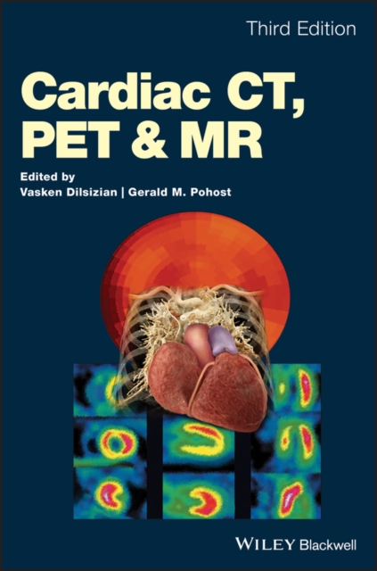
Cardiac CT, PET and MR
A complete guide to non-invasive imaging techniques in cardiology
Today?s imaging technologies offer cardiologists more ways than ever to diagnose conditions of the heart without the need of endoscopies and other invasive procedures. Now in its third edition,Cardiac CT, PET and MRI continues to provide an in-depth explanation of these tools and their correct applications, while also exploring cardiac imaging?s most recent and groundbreaking developments.
This wide-ranging guide places CT, PET and MRI in a practical context, illustrating clearly their respective functions as they apply to specific cardiological disorders and clinical situations. With the addition of seven new chapters, it also offers an expanded insight into PET – an increasingly popular and affordable diagnostic utility, hitherto underexplored in texts devoted to imaging. Cardiac CT, PET and MRI includes:
* Clinically focused examinations of CT, PET and MRI – the three most popular non-invasive imaging modalities
* Illustrative full-color photos and images
* Access to a companion website featuring additional content
Cardiologists, radiologists, nuclear medicine physicians, physicists, and imaging technologists alike will find the third edition of Cardiac CT, PET and MRI an informative and accessible resource with a direct use in their day-to-day practice.
eBook
Incremental value of left atrial booster and reservoir strain in predicting atrial fibrillation in patients with hypertrophic cardiomyopathy: a cardiovascular magnetic resonance study
Background
Left atrial (LA) size and function are known predictors of new onset atrial fibrillation (AF) in hypertrophic cardiomyopathy (HCM) patients. Components of LA deformation including reservoir, conduit, and booster function provide additional information on atrial mechanics. Whether or not LA deformation can augment our ability to predict the risk of new onset AF in HCM patients beyond standard measurements is unknown.
Methods
We assessed LA size, function, and deformation on cardiovascular magnetic resonance (CMR) in 238 genotyped HCM patients and compared this with twenty age, sex, blood pressure and body mass index matched control subjects. We further evaluated the determinants of new onset AF in HCM patients.
Results
Compared to control subjects, HCM patients had higher LA antero-posterior diameter, lower LA ejection fraction and lower LA reservoir (19.9 [17.1, 22.2], 21.6 [19.9, 22.9],
P
= 0.047) and conduit strain (10.6 ± 4.4, 13.7 ± 3.3,
P
= 0.002). LA booster strain did not differ between healthy controls and HCM patients, but HCM patients who developed new onset AF (n = 33) had lower booster strain (7.6 ± 3.3, 9.5 ± 3.0,
P
= 0.001) than those that did not (n = 205). In separate multivariate models, age, LA ejection fraction, and LA booster and reservoir strain were each independent determinants of AF. Age ≥ 55 years was the strongest determinant (HR 6.62, 95% CI 2.79–15.70), followed by LA booster strain ≤ 8% (HR 3.69, 95% CI 1.81–7.52) and LA reservoir strain ≤ 18% (HR 2.56, 95% CI 1.24–5.27). Conventional markers of HCM phenotypic severity, age and sudden death risk factors were associated with LA strain components.
Conclusions
LA strain components are impaired in HCM and, together with age, independently predicted the risk of new onset AF. Increasing age and phenotypic severity were associated with LA strain abnormalities. Our findings suggest that the routine assessment of LA strain components and consideration of age could augment LA size in predicting risk of AF, and potentially guide prophylactic anticoagulation use in HCM.
Journal Article
Motion-corrected 3D whole-heart water-fat high-resolution late gadolinium enhancement cardiovascular magnetic resonance imaging
Background
Conventional 2D inversion recovery (IR) and phase sensitive inversion recovery (PSIR) late gadolinium enhancement (LGE) cardiovascular magnetic resonance (CMR) have been widely incorporated into routine CMR for the assessment of myocardial viability. However, reliable suppression of fat signal, and increased isotropic spatial resolution and volumetric coverage within a clinically feasible scan time remain a challenge. In order to address these challenges, this work proposes a highly efficient respiratory motion-corrected 3D whole-heart water/fat LGE imaging framework.
Methods
An accelerated IR-prepared 3D dual-echo acquisition and motion-corrected reconstruction framework for whole-heart water/fat LGE imaging was developed. The acquisition sequence includes 2D image navigators (iNAV), which are used to track the respiratory motion of the heart and enable 100% scan efficiency. Non-rigid motion information estimated from the 2D iNAVs and from the data itself is integrated into a high-dimensional patch-based undersampled reconstruction technique (HD-PROST), to produce high-resolution water/fat 3D LGE images. A cohort of 20 patients with known or suspected cardiovascular disease was scanned with the proposed 3D water/fat LGE approach. 3D water LGE images were compared to conventional breath-held 2D LGE images (2-chamber, 4-chamber and stack of short-axis views) in terms of image quality (1: full diagnostic to 4: non-diagnostic) and presence of LGE findings.
Results
Image quality was considered diagnostic in 18/20 datasets for both 2D and 3D LGE magnitude images, with comparable image quality scores (2D: 2.05 ± 0.72, 3D: 1.88 ± 0.90,
p
-value = 0.62) and overall agreement in LGE findings. Acquisition time for isotropic high-resolution (1.3mm
3
) water/fat LGE images was 8.0 ± 1.4 min (3-fold acceleration, 60–88 slices covering the whole heart), while 2D LGE images were acquired in 5.6 ± 2.2 min (12–18 slices, including pauses between breath-holds) albeit with a lower spatial resolution (1.40–1.75 mm in-plane × 8 mm slice thickness).
Conclusion
A novel framework for motion-corrected whole-heart 3D water/fat LGE imaging has been introduced. The method was validated in patients with known or suspected cardiovascular disease, showing good agreement with conventional breath-held 2D LGE imaging, but offering higher spatial resolution, improved volumetric coverage and good image quality from a free-breathing acquisition with 100% scan efficiency and predictable scan time.
Journal Article

169 Two-dimensional and four-dimensional velocity imaging quantified mitral regurgitation is associated with left ventricular remodelling post mitral valve surgery
IntroductionAccurate assessment of degenerative mitral regurgitation (MRegurg) by transthoracic echocardiography (TTE) can be challenging. Phase-contrast cardiovascular magnetic resonance (PCMR) imaging enables reliable quantitation of MRegurg volume, even in the presence of eccentric and multiple jets, highlighting its advantage in the evaluation of degenerative MRegurg. Consequently, several studies demonstrated discordance of MRegurg severity between PCMR and TTE, with PCMR showing superior correlation with outcomes. Assessment of MRegurg volume by Four-dimensional Flow (4DF) CMR has been shown to be feasible and reproducible. This novel 3-dimensional, free-breathing technique facilitates direct assessment of flow across all four valves simultaneously in one, simple acquisition. 4DF CMR can therefore offer numerous clinical advantages in the evaluation of degenerative MRegurg.PurposeTo determine whether 4DF CMR quantitation of degenerative MRegurg volume is predictive of post-operative left ventricular (LV) remodelling.MethodsThis prospective, single centre, observational cohort study recruited 30 patients with degenerative MRegurg undergoing mitral valve surgery (repair/replacement). All patients underwent TTE, PCMR and 4DF CMR pre-surgery and a follow up PCMR at 6 months post-surgery. MRegurg volume was quantified by TTE (PISA method), PCMR (indirect method) and 4DF CMR (mitral inflow-aortic outflow method). Linear regression analysis was used to assess the predictive value of pre-operative MRegurg volume by these modalities with the post-operative change in the LV end-diastolic volume.ResultsMedian age was 69 (63-72) years; male n=21 (70%). The most common aetiology of MR was posterior mitral valve prolapse n=19 (63.3%), with a flail leaflet present in 10 patients (33.3%). Bland-Altman plots demonstrated smaller bias and narrower limits of agreement between MRegurg volume quantified by 4DF CMR and PCMR, than by the other methods (Figure 1). Furthermore, linear regression analysis demonstrated a significant association between the MRegurg volume and the post-operative LV remodelling, when the regurgitant volume was quantified by PCMR (p<0.001) and 4DF CMR (p=0.011), but not when assessed by TTE (p=0.518) (Figure 2).ConclusionThis study demonstrates the potential clinical utility of 4DF CMR in the assessment of degenerative mitral regurgitation.Abstract 169 Figure 1Bland-Altman plots Panel (A) presents Bland-Altman plot of relationship between mitral regurgitant volume quantified by 4DF CMR and PCMR, panel (B) by 4DF CMR and TTE-PISA and panel (C) PCMR and TTE-PISA. Green line represents bias, which is much smaller between 4DF CMR and PCMR (-11.61), than between 4DF CMR and TTE-PISA (98.39) and PCMR and TTE-PISA (102.99) 4DF CMR=4D flow cardiovascular magnetic resonance imaging; LLA=lower limit of agreement; PCMR=phase-contrast magnetic resonance imaging; RV=regurgitant volume; TTE-PISA=transthoracic echocardiography-proximal isovelocity surface area method; ULA=upper limit of agreementAbstract 169 Figure 2Linear regression analysis between post-operative change in left ventricular end-diastolic volume and pre-operative mitral regurgitant volume Panel (A) presents MR regurgitant volume quantified by 4DF CMR, panel (B) by PCMR and panel (C) by the TTE-PISA method 4DF CMR=4D flow cardiovascular magnetic resonance imaging; LVEDV=left ventricular end-diastolic volume; MR=mitral regurgitation; PCMR=phase-contrast magnetic resonance imaging; RVOL=regurgitant volume; TTE-PISA=transthoracic echocardiography-proximal isovelocity surface area methodConflict of InterestNone
Journal Article