Catalogue Search | MBRL


Are you sure you want to remove the book from the shelf?

{{itemTitle}}
1,786
result(s) for
"fiber pathways"
Sort by:

Prevalence of white matter pathways coming into a single white matter voxel orientation: The bottleneck issue in tractography
Characterizing and understanding the limitations of diffusion MRI fiber tractography is a prerequisite for methodological advances and innovations which will allow these techniques to accurately map the connections of the human brain. The so‐called “crossing fiber problem” has received tremendous attention and has continuously triggered the community to develop novel approaches for disentangling distinctly oriented fiber populations. Perhaps an even greater challenge occurs when multiple white matter bundles converge within a single voxel, or throughout a single brain region, and share the same parallel orientation, before diverging and continuing towards their final cortical or sub‐cortical terminations. These so‐called “bottleneck” regions contribute to the ill‐posed nature of the tractography process, and lead to both false positive and false negative estimated connections. Yet, as opposed to the extent of crossing fibers, a thorough characterization of bottleneck regions has not been performed. The aim of this study is to quantify the prevalence of bottleneck regions. To do this, we use diffusion tractography to segment known white matter bundles of the brain, and assign each bundle to voxels they pass through and to specific orientations within those voxels (i.e. fixels). We demonstrate that bottlenecks occur in greater than 50‐70% of fixels in the white matter of the human brain. We find that all projection, association, and commissural fibers contribute to, and are affected by, this phenomenon, and show that even regions traditionally considered “single fiber voxels” often contain multiple fiber populations. Together, this study shows that a majority of white matter presents bottlenecks for tractography which may lead to incorrect or erroneous estimates of brain connectivity or quantitative tractography (i.e., tractometry), and underscores the need for a paradigm shift in the process of tractography and bundle segmentation for studying the fiber pathways of the human brain. \"Bottleneck\" pose a challenge for diffusion tractography, and hinder our ability to accurately map the structural connections of the brain. We demonstrate that bottlenecks occur in greater than 50–70% of fixels in the human brain white matter, and find that all projection, association, and commissural fibers contribute to, and are affected by, this phenomenon. These results emphasize the use of caution when interpreting quantitative diffusion magnetic resonance imaging connectomics results, and underscore the need for a paradigm shift in the process of tractography for studying the fiber pathways of the human brain.
Journal Article

Tractography dissection variability: What happens when 42 groups dissect 14 white matter bundles on the same dataset?
White matter bundle segmentation using diffusion MRI fiber tractography has become the method of choice to identify white matter fiber pathways in vivo in human brains. However, like other analyses of complex data, there is considerable variability in segmentation protocols and techniques. This can result in different reconstructions of the same intended white matter pathways, which directly affects tractography results, quantification, and interpretation. In this study, we aim to evaluate and quantify the variability that arises from different protocols for bundle segmentation. Through an open call to users of fiber tractography, including anatomists, clinicians, and algorithm developers, 42 independent teams were given processed sets of human whole-brain streamlines and asked to segment 14 white matter fascicles on six subjects. In total, we received 57 different bundle segmentation protocols, which enabled detailed volume-based and streamline-based analyses of agreement and disagreement among protocols for each fiber pathway. Results show that even when given the exact same sets of underlying streamlines, the variability across protocols for bundle segmentation is greater than all other sources of variability in the virtual dissection process, including variability within protocols and variability across subjects. In order to foster the use of tractography bundle dissection in routine clinical settings, and as a fundamental analytical tool, future endeavors must aim to resolve and reduce this heterogeneity. Although external validation is needed to verify the anatomical accuracy of bundle dissections, reducing heterogeneity is a step towards reproducible research and may be achieved through the use of standard nomenclature and definitions of white matter bundles and well-chosen constraints and decisions in the dissection process.
Journal Article

Inhibition of hippocampal mossy fiber plasticity and episodic memory by human Aβ oligomers is prevented by enhancing cAMP signaling in Alzheimer's mice
INTRODUCTION Early episodic memory impairment in Alzheimer's disease (AD) is linked to synaptic dysfunction from amyloid β‐protein oligomers (oAβ), particularly affecting the dentate gyrus mossy fiber‐CA3 pathway. The APPNL‐G‐F mouse model exhibits early deficits in mossy fiber long‐term potentiation (mf‐LTP). METHODS We administered the β‐adrenergic receptor agonist isoproterenol (ISO) in vivo and phosphodiesterase type 4 inhibitor GSK356278 in vitro to assess their impact on mf‐LTP and contextual fear memory. Fluorescence lifetime imaging (FLIM)‐Förster resonance energy transfer (FRET) microscopy was used to visualize impaired and rescued cyclic adenosine monophosphate (cAMP) signaling in dentate gyrus neurons. RESULTS ISO prevented mf‐LTP impairment at 3–4 mo and improved memory by 7 mo. GSK356278 inhibited mf‐LTP deficits in a dose‐dependent manner. ISO also reduced hyperphosphorylation of synapsin I and microgliosis. DISCUSSION These findings suggest that β‐AR activation and phosphodiesterase 4 (PDE4) inhibition mitigate oAβ‐induced memory deficits, supporting enhanced cAMP signaling as a therapeutic target for early AD. Highlights Early episodic memory deficits in AD linked to oAβ‐induced synaptic dysfunction. Isoproterenol and GSK356278 improve mossy fiber‐LTP and fear memory deficits. FLIM‐FRET shows treatments restore cAMP signaling in dentate gyrus neurons. Isoproterenol reduces synapsin I hyperphosphorylation and microgliosis. Enhancing cAMP signaling may help mitigate early memory deficits in AD.
Journal Article
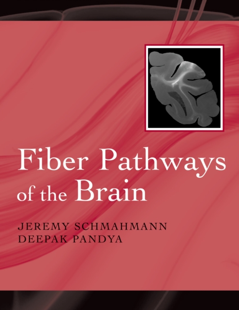
Fiber pathways of the brain
Beautifully illustrated, this unique volume is a comprehensive study of the corticocortical and corticosubocortical connections in the cerebral cortex of the rhesus monkey.It will give readers a detailed understanding of the white matter fiber pathways of the brain.
eBook
Serial optical coherence scanner for large-scale brain imaging at microscopic resolution
We describe a serial optical coherence scanner (SOCS) for high resolution imaging of ex-vivo brain. SOCS integrates a multi-contrast optical coherence tomography and a vibratome slicer to establish comprehensive brain anatomy and fiber pathways in three-dimensional space. Rat brain images are demonstrated by utilizing intrinsic optical contrasts including back-scattering, birefringence and optic axis orientation, which are simultaneously generated from the same dataset. Volumetric images from serial scans are combined to realize large scale brain maps. Nerve fiber tracts are globally described in 3D by retardance, and delicately delineated by cross-polarization at the resolution of 15×15×5.5μm3. In-plane orientations of the tracts are quantified by optic axis orientation. SOCS offers a new solution for complete reconstructions of macroscopic tissues such as primate and human brains at microscopic resolution. The technique also opens up varieties of opportunities for connectome studies and systematic investigations on neurological diseases and brain disorders.
•Serial optical coherence scanner enables large-scale high-resolution tissue imaging.•Ex-vivo rat brain is reconstructed and global neural roadmaps are depicted in 3D.•Trajectories of fiber tracts are visualized at the resolution of 15×15×5.5μm3.•Fiber orientation is quantified with optical measurement.
Journal Article

Scatterometry Measurements With Scattered Light Imaging Enable New Insights Into the Nerve Fiber Architecture of the Brain
The correct reconstruction of individual (crossing) nerve fibers is a prerequisite when constructing a detailed network model of the brain. The recently developed technique Scattered Light Imaging (SLI) allows the reconstruction of crossing nerve fiber pathways in whole brain tissue samples with micrometer resolution: the individual fiber orientations are determined by illuminating unstained histological brain sections from different directions, measuring the transmitted scattered light under normal incidence, and studying the light intensity profiles of each pixel in the resulting image series. So far, SLI measurements were performed with a fixed polar angle of illumination and a small number of illumination directions, providing only an estimate of the nerve fiber directions and limited information about the underlying tissue structure. Here, we use a display with individually controllable light-emitting diodes to measure the full distribution of scattered light behind the sample (scattering pattern) for each image pixel at once, enabling scatterometry measurements of whole brain tissue samples. We compare our results to coherent Fourier scatterometry (raster-scanning the sample with a non-focused laser beam) and previous SLI measurements with fixed polar angle of illumination, using sections from a vervet monkey brain and human optic tracts. Finally, we present SLI scatterometry measurements of a human brain section with 3 μm in-plane resolution, demonstrating that the technique is a powerful approach to gain new insights into the nerve fiber architecture of the human brain.
Journal Article
White matter development during late adolescence in healthy males: A cross-sectional diffusion tensor imaging study
Previous MRI studies of healthy children have reported age-related white matter (WM) changes in language and motor areas of the brain. The authors investigated WM development in healthy adolescent males through age-associated changes in fractional anisotropy (FA), radial (λ⊥) and axial (λ||) diffusivity.
Twenty-four healthy adolescent males (mean age=16.6, SD=2.5 years) were divided into two groups with an age split of 16.9 years and underwent a whole-brain voxelwise analysis.
At a threshold of p<0.001 and extent threshold of 100 contiguous voxels, several clusters with increased FA and axial diffusivity and no differences in radial diffusivity were observed in older adolescents compared to the younger adolescents in the left arcuate fasciculus, bilateral posterior internal capsule/thalamic radiation, bilateral prefrontal gyrus, right superior temporal gyrus, and posterior corpus callosum. Increased FA and λ|| of several clusters along the arcuate fasciculus significantly correlated with a test of language and semantic memory.
These results suggest ongoing maturational changes especially in the arcuate fasiculus during late adolescence. Increased FA and λ|| with no changes in radial diffusivity may reflect a developmental pattern of reduced tortuousity toward more straightened fibers and/or increased axonal fiber organization during late adolescence.
Journal Article

A mathematical model of ephaptic interactions in neuronal fiber pathways: Could there be more than transmission along the tracts?
While numerous studies of ephaptic interactions have focused on either axons of peripheral nerves or on cortical structures, no attention has been given to the possibility of ephaptic interactions in white matter tracts. Inspired by the highly organized, tightly packed geometry of axons in fiber pathways, we aim to investigate the potential effects of ephaptic interactions along these structures that are resilient to experimental probing. We use axonal cable theory to derive a minimal model of a sheet of
ephaptically coupled axons. Numerical solutions of the proposed model are explored as ephaptic coupling is varied. We demonstrate that ephaptic interactions can lead to local phase locking between adjacent traveling impulses and that, as coupling is increased, traveling impulses trigger new impulses along adjacent axons, resulting in finite size traveling fronts. For strong enough coupling, impulses propagate laterally and backwards, resulting in complex spatiotemporal patterns. While common large-scale brain network models often model fiber pathways as simple relays of signals between different brain regions, our work calls for a closer reexamination of the validity of such a view. The results suggest that in the presence of significant ephaptic interactions, the brain fiber tracts can act as a dynamic active medium.
Starting from the FitzHugh-Nagumo cable model, we derive a system of nonlinear coupled partial differential equations (PDEs) to model a sheet of
ephaptically coupled axons. We also present a continuous limit approximation transforming the model into a two-dimensional field equation. We numerically solve the equations exploring the dynamics as coupling strength is varied. We observe phase locking of adjacent impulses and coordination of subthreshold dynamics. Strong enough coupling generates complex spatiotemporal patterns as new impulses form traveling fronts propagating laterally and backwards. The transition between different dynamic regimes happens abruptly at critical values of parameter. The results put into question the validity of assuming the role of fiber pathways to be that of mere interneuronal transmission and call for further investigation of the matter.
Journal Article

Both Nuclear and Membrane Estrogen Receptor Alpha Impact the Expression of Estrogen Receptors and Plasticity Markers in the Mouse Hypothalamus and Hippocampus
Estrogens via estrogen receptor alpha (ERα) genomic and nongenomic signaling can influence plasticity processes in numerous brain regions. Using mice that express nuclear only ERα (NOER) or membrane only ERα (MOER), this study examined the effect of receptor compartmentalization on the paraventricular nucleus of the hypothalamus (PVN) and the hippocampus. The absence of nuclear and membrane ERα expression impacted females but not males in these two brain areas. In the PVN, quantitative immunohistochemistry showed that the absence of nuclear ERα increased nuclear ERβ. Moreover, in the hippocampus CA1, immuno-electron microscopy revealed that the absence of either nuclear or membrane ERα decreased extranuclear ERα and pTrkB in synapses. In contrast, in the dentate gyrus, the absence of nuclear ERα increased pTrkB in synapses, whereas the absence of membrane ERα decreased pTrkB in axons. However, the absence of membrane only ERα decreased the sprouting of mossy fibers in CA3 as reflected by changes in zinc transporter immunolabeling. Altogether these findings support the idea that both membrane and nuclear ERα contribute overlapping and unique actions of estrogen that are tissue- and cellular-specific.
Journal Article

Single-digit arithmetic processing-anatomical evidence from statistical voxel-based lesion analysis
Different specific mechanisms have been suggested for solving single-digit arithmetic operations. However, the neural correlates underlying basic arithmetic (multiplication, addition, subtraction) are still under debate. In the present study, we systematically assessed single-digit arithmetic in a group of acute stroke patients (n = 45) with circumscribed left- or right-hemispheric brain lesions. Lesion sites significantly related to impaired performance were found only in the left-hemisphere damaged (LHD) group. Deficits in multiplication and addition were related to subcortical/white matter brain regions differing from those for subtraction tasks, corroborating the notion of distinct processing pathways for different arithmetic tasks. Additionally, our results further point to the importance of investigating fiber pathways in numerical cognition.
Journal Article