Catalogue Search | MBRL


Are you sure you want to remove the book from the shelf?

{{itemTitle}}
10
result(s) for
"postmortem CT-angiography"
Sort by:

Development of a protocol for standardized use of a water-soluble contrast agent with polyethylene glycol in post-mortem CT angiography
Computed tomography angiography (PMCTA) is increasingly used in postmortem cases. Standardized validated protocols permit to compare different PMCTA images and make it more easily to defend a case in court. In addition to the well-known technique by Grabherr et al. (2011) which is using paraffin oil as a carrier substance, water-soluble polyethylene glycol 200 (PEG200) can be used in combination with the contrast agent Accupaque® 300. As to date, there exists no standardized protocol for the use of this contrast agent mixture, the aim of this study was to develop a protocol using it. Between 2012 and 2022, 23 PMCTA with PEG200 and Accupaque®300 were performed at the University Centre of Legal Medicine Lausanne (Switzerland) and the Institute of Forensic Medicine Munich (Germany). The images obtained were evaluated regarding the opacification of the vessels and possible artefacts. The best image quality was obtained with a mixing ratio of 1:15 (Accupaque®300:PEG200) and a perfusion volume of 1000 ml in the arterial, 1400 ml in the venous and 350 ml in the dynamic phase. The infusion rates described by Grabherr et al. were confirmed for the three phases. Overall, the opacification of the vessels was diagnostically sufficient. In 13 cases no opacification of the right coronary artery was observed due to a stratification artefact. By using the PMCTA protocol with PEG200 as a carrier, a good overall image quality can be achieved. This protocol offers the possibility to standardize PMCTA with PEG200.
Journal Article
Postmortem imaging of sudden cardiac death
Postmortem imaging is increasingly used in forensic practice in cases of natural deaths related to cardiovascular diseases, which represent the most common causes of death in developed countries. While radiological examination is generally considered to be a good complement for conventional autopsy, it was thought to have limited application in cardiovascular pathology. At present, multidetector computed tomography (MDCT), CT angiography, and cardiac magnetic resonance imaging (MRI) are used in postmortem radiological investigation of cardiovascular pathologies. This review presents the actual state of postmortem imaging for cardiovascular pathologies in cases of sudden cardiac death (SCD), taking into consideration both the advantages and limitations. The radiological evaluation of ischemic heart disease (IHD), the most frequent cause of SCD in the general population of industrialized countries, includes the examination of the coronary arteries and myocardium. Postmortem CT angiography (PMCTA) is very useful for the detection of stenoses and occlusions of coronary arteries but less so for the identification of ischemic myocardium. MRI is the method of choice for the radiological investigation of the myocardium in clinical practice, but its accessibility and application are still limited in postmortem practice. There are very few reports implicating postmortem radiology in the investigation of other causes of SCD, such as cardiomyopathies, coronary artery abnormalities, and valvular pathologies. Cardiomyopathies representing the most frequent cause of SCD in young athletes cannot be diagnosed by echocardiography, the most widely available technique in clinical practice for the functional evaluation of the heart and the detection of cardiomyopathies. PMCTA and MRI have the potential to detect advanced stages of diseases when morphological substrate is present, but these methods have yet to be sufficiently validated for postmortem cases. Genetically determined channelopathies cannot be detected radiologically. This review underlines the need to establish the role of postmortem radiology in the diagnosis of SCD.
Journal Article

Postmortem imaging as a complementary tool for the investigation of cardiac death
In the past 2 decades, modern radiological methods, such as multiple detector computed tomography (MDCT), MDCT-angiography, and cardiac magnetic resonance imaging (MRI) were introduced into postmortem practice for investigation of sudden death (SD), including cases of sudden cardiac death (SCD). In forensic cases, the underlying cause of SD is most frequently cardiovascular with coronary atherosclerotic disease as the leading cause. There are many controversies about the role of postmortem imaging in establishing the cause of death and especially the value of minimally invasive autopsy techniques. This paper discusses the state of the art for postmortem radiological evaluation of the heart compared to classical postmortem examination, especially in cases of SCD. In SCD cases, postmortem CT is helpful to estimate the heart size and to visualize haemopericardium and calcified plaques and valves, as well as to identify and locate cardiovascular devices. Angiographic methods are useful to provide a detailed view of the coronary arteries and to analyse them, especially regarding the extent and location of stenosis and obstruction. In postsurgical cases, it allows verification and documentation of the patency of stents and bypass grafts before opening the body. Postmortem MRI is used to investigate soft tissues such as the myocardium, but images are susceptible to postmortem changes and further work is necessary to increase the understanding of these radiological aspects, especially of the ischemic myocardium. In postsurgery cases, the value of postmortem imaging of the heart is reportedly for the diagnostic and documentation purposes. The implementation of new imaging methods into routine postmortem practice is challenging, as it requires not only an investment in equipment but, more importantly, investment in the expertise of interpreting the images. Once those requirements are implemented, however, they bring great advantages in investigating cases of SCD, as they allow documentation of the body, orientation of sampling for further analyses and gathering of other information that cannot be obtained by conventional autopsy such as a complete visualization of the vascular system using postmortem angiography.
Key points
There are no established guidelines for the interpretation of postmortem imaging examination of the heart
At present, postmortem imaging methods are considered as less accurate than the autopsy for cardiac deaths
Postmortem imaging is useful as a complementary tool for cardiac deaths
There is still a need to validate postmortem imaging in cardiac deaths by comparing with autopsy findings
Journal Article
Pathomorphological and CT-angiographical characteristics of coronary atherosclerotic plaques in cases of sudden cardiac death
The goal of this study was to assess the localization and types of thrombosed plaques in cases of sudden cardiac death attributed to coronary artery disease and to evaluate possible correlations with body mass index (BMI) and increased heart weight. This retrospective study was performed on forensic cases for which the cause of death was attributed to coronary artery disease. A complete autopsy and a multi-phase postmortem computed tomography (CT) angiography (MPMCTA) were performed in all cases. Eighty-five cases were selected (mean age, 55.18 ± 11.04 years; 72 men and 13 women). MPMCTA performed prior to autopsy enabled an evaluation of coronary artery perfusion before dissection of the body and helped therefore to guide sampling for histology. An acute coronary thrombosis was found in 57 cases, which included plaque erosion in 26 cases (mean age, 46.73 ± 8.33 years) and rupture or intra-plaque hemorrhage in 31 cases (mean age, 58.23 ± 10.62 years). Erosions were most frequently found in the left anterior descending artery (61.5 %), while only 35.48 % of ruptures were observed in this artery. Chronic coronary pathology was considered as the main cause of death in 28 cases (mean age, 59.64 ± 9.47 years). Sixty-two of the cases (72.94 %) had a BMI in the overweight category (BMI ≥25), with the highest mean BMI in patients with chronic coronary pathology without acute thrombosis found at autopsy. The heart weight was above the predicted reference values in 52 cases (61.18 %). Our results are in accordance with previously published studies on the spatial distribution of vulnerable plaques. We observed a higher percentage of eroded plaques than previously reported. Patients with coronary erosions were significantly younger than those with plaque rupture or those without an acute coronary thrombosis (
p
values <0.0001). BMI and heart weight were significantly higher for cases without thrombosis in comparison with those with plaque rupture (
p
values 0.028 and 0.003, respectively). Our results indicating that increased BMI and overweight hearts are associated with chronic ischemic heart disease are compatible with clinical studies. Performing more postmortem studies on forensic autopsies, including modern radiological examinations with MPMCTA, can enhance the detection of vulnerable plaques in living patients and prevent sudden cardiac death.
Journal Article

Postmortale bildgebende Verfahren
Obduktionen sind ein Schlüssel zu einem besseren Verständnis der Anatomie, (Patho‑)Physiologie und der (Patho‑)Morphologie. In der Rechtsmedizin ist die virtuelle Autopsie seit den späten 1990ern ein Standardinstrument der Obduktionstechnik. Vorteile der postmortalen Bildgebung sind die Erfassung des Ist-Zustands der Leiche von Kopf bis Fuß, mit exzellenter Beurteilbarkeit von Pathologien des Skeletts, Lufteinschlüssen und bei Durchführung einer Angiographie auch sehr gute Beurteilbarkeit des Herz-Kreislauf-Systems. Schwächen der postmortalen Bildgebung sind der eingeschränkte Weichgewebskontrast im CT, das Fehlen der haptischen Befunde, der Farbveränderungen, des Geruchs, die eingeschränkte Verfügbarkeit der CT und insbesondere der MRT sowie die für die Auswertung der radiologischen Daten notwendige Erfahrung mit postmortaler Bildgebung. Die postmortale Bildgebung kann bislang die klinisch-pathologische Obduktion nur ergänzen und keinesfalls ersetzen. Nichtsdestotrotz bereichert die postmortale Bildgebung die klinisch-pathologische Obduktion um neue technische Möglichkeiten. Inwieweit es gelingt, mit der postmortalen Bildgebung, insbesondere auch mit ergänzenden CT- oder MRT-gesteuerten Probebiopsien bzgl. der Todesursachenfeststellung valide Ergebnisse zu erzielen, muss systematisch evaluiert werden. Der vorliegende Artikel soll Pathologen einen Überblick über den Stand der Technik vermitteln und dazu anregen, die Technik aus der Pathologie heraus für pathologiespezifische Fragestellungen weiter zu entwickeln.
Journal Article
A calcification subtraction method for postmortem coronary computed tomography angiography
Although coronary computed tomography (CT) angiography is a useful tool for evaluating coronary artery lesions both ante- and postmortem, accurate evaluation of the lumen is difficult when highly calcified lesions are present, owing to overestimation of stenosis caused by blooming and partial volume artifacts. In clinical practice, to overcome this diagnostic problem, a subtraction method has been devised to remove calcification by subtracting the precontrast image from the contrast image. In this report, we describe a calcification subtraction method using image analysis software for postmortem coronary CT angiography. This method was devised based on preliminary experimental results showing that the most accurate subtraction was achieved using images reconstructed with a narrower field of view and bone kernel, resulting in higher spatial resolution. This subtraction method allowed evaluation of lumen patency and the degree of stenosis on contrast-enhanced images in a verification using actual specimens where evaluation of the lumen had been difficult because of high calcification. The results were morphologically similar to the macroscopic findings. This method allows more rapid and reliable lesion retrieval and is expected to be useful for postmortem coronary angiography in forensic practice.
Journal Article
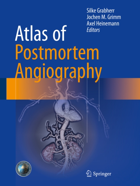
Atlas of postmortem angiography
This atlas of postmortem angiography provides a summary of techniques that have been developed and used in order to visualize the human vascular system.The indications, advantages, limitations, and pitfalls of the different techniques are explained in detail through the use of examples from real cases and a wealth of informative images, as well.
eBook
Impact of increasing levels of adaptive statistical iterative reconstruction on image quality in oil-based postmortem CT angiography in coronary arteries
IntroductionPostmortem multi-detector computed tomography (PMCT) has become an important part in forensic imaging. Modern reconstruction techniques such as iterative reconstruction (IR) are frequently used in postmortem CT angiography (PMCTA). The image quality of PMCTA depends on the strength of IR. For this purpose, we aimed to investigate the impact of different advanced IR levels on the objective and subjective PMCTA image quality.Material and methodsWe retrospectively analyzed the coronary arteries of 27 human cadavers undergoing whole-body postmortem CT angiography between July 2017 and March 2018 in a single center. Iterative reconstructions of the coronary arteries were processed in five different level settings (0%; 30%; 50%; 70%; 100%) by using an adaptive statistical IR method. We evaluated the objective (contrast-to-noise ratio (CNR)) and subjective image quality in several anatomical locations.ResultsOur results demonstrate that the increasing levels of an IR technique have relevant impact on the image quality in PMCTA scans in forensic postmortem examinations. Higher levels of IR have led to a significant reduction of image noise and therefore to a significant improvement of objective image quality (+ 70%). However, subjective image quality is inferior at higher levels of IR due to plasticized image appearance.ConclusionObjective image quality in PMCTA progressively improves with increasing level of IR with the best CNR at the highest IR level. However, subjective image quality is best at low to medium levels of IR. To obtain a “classic” image appearance with optimal image quality, PMCTAs should be reconstructed at medium levels of IR.
Journal Article

Computed Tomography: The Revolution in Postmortem Angiography
Multidetector computed tomography (MDCT) is the most widespread tool in modern forensic radiology. Thanks to a short examination time, high spatial resolution, and the possibility of reconstructions in different axes and two and three dimensions, it is an excellent method for rapidly examining a whole body and performing a first analysis of lesions. In clinical radiology, the sensitivity of MDCT can be increased by injecting contrast agent and enhancing the obtained images, leading to better visualization of organ tissues and of the vascular system. This clinical experience inspired researchers in postmortem imaging to start investigating the possibility of performing postmortem MDCT angiography. Similar to the clinical experience, the aim is to increase the sensitivity of postmortem computed tomography (PMCT) and allow investigation of the vascular system, which is often too complex to examine in conventional autopsy, especially if small vessels are to be investigated. However, the application of contrast agent postmortem is challenging and differs from the process in clinical angiography, principally because of the absence of cardiovascular circulation and postmortem changes in the body. Although there are multiple classic methods for performing postmortem angiography, they cannot simply be transferred to modern forensic imaging because they focus primarily on single organs or on embryos and fetuses. Therefore, to overcome these problems, different approaches have been developed for performing modern, minimally invasive PMCT angiography (PMCTA). Once such a technique is established, questions arise about how to interpret the images because they differ from those of clinical angiography. Additionally, the injection of contrast medium into a body that is associated with a medico-legal or forensic investigation implies the problem of eventual alteration of the results or further analyses that are normally performed in such cases. This chapter explains the meaning of PMCTA and the challenges encountered in establishing methods that are applicable in legal medicine; it gives a short overview of the most frequently used techniques in modern forensic imaging.
Book Chapter
Postmortem Whole Body CT Angiography Using Aqueous Contrast Agent
In modern postmortem imaging, the visualization of the vascular system by postmortem angiography is mostly done using multidetector computed tomography (CT). Like clinical radiologic investigations, whole body angiography makes the vascular system visible and allows identification of vascular lesions such as traumatic dissection or rupture. Additionally, the injection of contrast agent enhances soft tissue and aids in the visualization of lesions in organ parenchyma. In this chapter, we describe two methods of whole body postmortem CT angiography (PMCTA) that can be classed in the category of “PMCTA using aqueous contrast agent.”
Book Chapter