Catalogue Search | MBRL


Are you sure you want to remove the book from the shelf?

{{itemTitle}}
1,974
result(s) for
"Cat Diseases - pathology"
Sort by:

Pathogenesis of Dermatophytosis
Despite the superficial localization of most dermatophytosis, host-fungus relationship in these infections is complex and still poorly elucidated. Though many efforts have been accomplished to characterize secreted dermatophytic proteases at the molecular level, only punctual insights have been afforded into other aspects of the pathogenesis of dermatophytosis, such as fungal adhesion, regulation of gene expression during the infection process, and immunomodulation by fungal factors. However, new genetic tools were recently developed, allowing a more rapid and high-throughput functional investigation of dermatophyte genes and the identification of new putative virulence factors. In addition, sophisticated in vitro infection models are now used and will open the way to a more comprehensive view of the interactions between these fungi and host epidermal cells, especially keratinocytes.
Journal Article

Bartonella infections in cats and dogs including zoonotic aspects
Bartonellosis is a vector-borne zoonotic disease with worldwide distribution that can infect humans and a large number of mammals including small companion animals (cats and dogs). In recent years, an increasing number of studies from around the world have reported
Bartonella
infections, although publications have predominantly focused on the North American perspective. Currently, clinico-pathological data from Europe are more limited, suggesting that bartonellosis may be an infrequent or underdiagnosed infectious disease in cats and dogs. Research is needed to confirm or exclude
Bartonella
infection as a cause of a spectrum of feline and canine diseases.
Bartonella
spp. can cause acute or chronic infections in cats, dogs and humans. On a comparative medical basis, different clinical manifestations, such as periods of intermittent fever, granulomatous inflammation involving the heart, liver, lymph nodes and other tissues, endocarditis, bacillary angiomatosis, peliosis hepatis, uveitis and vasoproliferative tumors have been reported in cats, dogs and humans. The purpose of this review is to provide an update and European perspective on
Bartonella
infections in cats and dogs, including clinical, diagnostic, epidemiological, pathological, treatment and zoonotic aspects.
Journal Article

SARS-CoV-2 infection, disease and transmission in domestic cats
Severe Acute Respiratory Syndrome Coronavirus 2 (SARS-CoV-2) is the cause of Coronavirus Disease 2019 (COVID-19) and responsible for the current pandemic. Recent SARS-CoV-2 susceptibility studies in cats show that the virus can replicate in these companion animals and transmit to other cats. Here, we present an in-depth study of SARS-CoV-2 infection, disease and transmission in domestic cats. Cats were challenged with SARS-CoV-2 via intranasal and oral routes. One day post challenge (DPC), two sentinel cats were introduced. Animals were monitored for clinical signs, clinicopathological abnormalities and viral shedding. Postmortem examinations were performed at 4, 7 and 21 DPC. Viral RNA was not detected in blood but transiently in nasal, oropharyngeal and rectal swabs and bronchoalveolar lavage fluid as well as various tissues. Tracheobronchoadenitis of submucosal glands with the presence of viral RNA and antigen was observed in airways of the infected cats. Serology showed that both, principals and sentinels, developed antibodies to SARS-CoV-2. All animals were clinically asymptomatic during the course of the study and capable of transmitting SARS-CoV-2 to sentinels. The results of this study are critical for understanding the clinical course of SARS-CoV-2 in a naturally susceptible host species, and for risk assessment.
Journal Article

Invasive Tracheal and Cranial Mediastinal Aspergillosis in a Young Otherwise Healthy Cat
Abstract
A 3.5-year-old castrated male domestic medium hair cat was evaluated for dry cough and labored breathing. A cranial mediastinal mass was seen on thoracic radiographs. On computed tomography, the mass displaced the cranial vena cava and dorsally displaced and compressed the intrathoracic trachea. The patient was taken to surgery for attempted mass removal. Intraoperatively, the mass was adhered to the cranial vena cava, aortic arch, left subclavian artery, and had partially engulfed the brachiocephalic trunk. The cat was euthanized and on necropsy the mass was found to invade the lumen of the vena cava and the tracheal lumen. An Aspergillus lentulus fungal granuloma was diagnosed histologically and by fungal culture and PCR. We highlight the difficulty in determining the extent of invasion with invasive aspergillosis and provide evidence that invasive aspergillosis can occur in otherwise healthy, young cats with no concurrent immunosuppressive treatments or comorbidities.
Journal Article
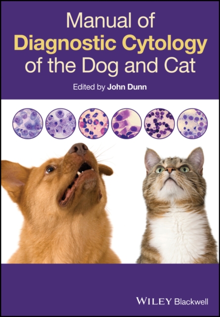
Manual of Diagnostic Cytology of the Dog and Cat
Wish you could interpret cytological specimens in practice rather than paying a lab to do it for you? Want to provide your clients with a faster service?
Manual of Diagnostic Cytology of the Dog and Cat is the ideal quick reference for the busy veterinarian in first opinion practice. It describes techniques for obtaining good quality cytological diagnostic specimens, and guides you through the interpretation of cytological findings.
Created to be used alongside the microscope, hundreds of high quality colour photos will help you to identify normal cell types and abnormal cytology, including both non-neoplastic and neoplastic lesions. It describes in a clear and concise manner the most common lesions and related disorders encountered in a practice setting. The concise format means that you can quickly find exactly what you're looking for.
Covering indications for cytological investigation, collection techniques and the evaluation and interpretation of findings, this concise manual will be your go-to resource.
eBook

Neutralizing antibodies for SARS-CoV-2 in stray animals from Rio de Janeiro, Brazil
The epidemic of coronavirus disease 2019 (COVID-19), caused by a novel Betacoronavirus named severe acute respiratory syndrome coronavirus 2 (SARS-CoV-2) became a public health emergency worldwide. Few reports indicate that owned pets from households with at least one human resident that was diagnosed with COVID-19 can be infected by SARS-CoV-2. However, the exposure to SARS-CoV-2 of pets from households with no COVID-19 cases or stray animals remains less assessed. Using real-time reverse transcriptase polymerase chain reaction (RT-PCR) and plaque reduction neutralization test (PRNT 90 ), we investigated the infection and previous exposure of dogs and cats to SARS-CoV-2 during the ongoing COVID-19 epidemic in Rio de Janeiro, Brazil. From June to August 2020, 96 animals were sampled, including 49 cats (40 owned and 9 stray) and 47 dogs (42 owned and 5 stray). Regarding owned pets, 75.6% (62/82) belonged to households with no COVID-19 cases. Samples included serum, and rectal and oropharyngeal swabs. All swabs were negative for SARS-CoV-2 RNA, but serum samples of a stray cat and a stray dog presented neutralizing antibodies for SARS-CoV-2, with PRNT 90 titer of 80 and 40, respectively. Serological data presented here suggest that not only owned pets from households with COVID19 cases, but also stray animals are being exposed to SARS-CoV-2 during the COVID-19 pandemic.
Journal Article
Short colon syndrome in cats
Abstract
Background
Shortening of the colon has been described in cats, but its imaging and clinicopathological features remain poorly understood.
Objectives
Description of the signalment, clinical presentation, imaging, endoscopic and histological features of short colon syndrome in cats.
Animals
Ninety-three cats diagnosed with short colon.
Methods
Multi-institutional, descriptive, retrospective case series study. Medical records were searched for a diagnosis of short colon on abdominal ultrasonography, computed tomography, endoscopy, autopsy, or a combination of these modalities.
Results
The median age of included cats was 12 years at the time of diagnosis. Diarrhea was the most common clinical sign (60/92; 65%), followed by vomiting (36/92; 39%), weight loss (36/92; 39%), and inappetence (24/92; 26%). Thirteen percent of cats (12/92) had no signs of gastrointestinal disease at the time of diagnosis. In addition to a shortened colonic length, 79% (66/84) of cats had concomitant colonic thickening on ultrasonographic examination. On colonoscopy, mucosal ulcerations of the colonic wall were seen in 39% (9/23) of cats. Histopathologically, all cats but 1 (diagnosed simultaneously with colonic small cell lymphoma) had lymphoplasmacytic colitis, and when small intestinal biopsies were performed, concurrent lymphoplasmacytic enteritis or small cell lymphoma of the small intestine.
Conclusions and Clinical Importance
Lymphoplasmacytic colitis is seen commonly in cats with short colon, suggesting a potential link between these entities.
Journal Article

Feline Calicivirus Virulent Systemic Disease: Clinical Epidemiology, Analysis of Viral Isolates and In Vitro Efficacy of Novel Antivirals in Australian Outbreaks
Feline calicivirus (FCV) causes upper respiratory tract disease (URTD) and sporadic outbreaks of virulent systemic disease (FCV-VSD). The basis for the increased pathogenicity of FCV-VSD viruses is incompletely understood, and antivirals for FCV-VSD have yet to be developed. We investigated the clinicoepidemiology and viral features of three FCV-VSD outbreaks in Australia and evaluated the in vitro efficacy of nitazoxanide (NTZ), 2′-C-methylcytidine (2CMC) and NITD-008 against FCV-VSD viruses. Overall mortality among 23 cases of FCV-VSD was 39%. Metagenomic sequencing identified five genetically distinct FCV lineages within the three outbreaks, all seemingly evolving in situ in Australia. Notably, no mutations that clearly distinguished FCV-URTD from FCV-VSD phenotypes were identified. One FCV-URTD strain likely originated from a recombination event. Analysis of seven amino-acid residues from the hypervariable E region of the capsid in the cultured viruses did not support the contention that properties of these residues can reliably differentiate between the two pathotypes. On plaque reduction assays, dose–response inhibition of FCV-VSD was obtained with all antivirals at low micromolar concentrations; NTZ EC50, 0.4–0.6 µM, TI = 21; 2CMC EC50, 2.7–5.3 µM, TI > 18; NITD-008, 0.5 to 0.9 µM, TI > 111. Investigation of these antivirals for the treatment of FCV-VSD is warranted.
Journal Article
Cats shedding pathogenic Leptospira spp.—An underestimated zoonotic risk?
Shedding of DNA of pathogenic Leptospira spp. has been documented in naturally infected cats in several countries, but urinary shedding of infectious Leptospira spp. has only recently been proven. The climate in Southern Chile is temperate rainy with high annual precipitations which represents ideal preconditions for survival of Leptospira spp., especially during spring and summer. The aims of this study were to investigate shedding of pathogenic Leptospira spp. in outdoor cats in Southern Chile, to perform molecular characterization of isolates growing in culture, and to assess potential risk factors associated with shedding. Urine samples of 231 outdoor cats from rural and urban areas in southern Chile were collected. Urine samples were investigated for pathogenic Leptospira spp. by 4 techniques: qPCR targeting the lipL32 gene, immunomagnetic separation (IMS)-coupled qPCR (IMS-qPCR), direct culture and IMS-coupled culture. Positive urine cultures were additionally confirmed by PCR. Multilocus sequence typing (MLST) was used to molecularly characterize isolates obtained from positive cultures. Overall, 36 urine samples (15.6%, 95% confidence interval (CI) 11.4-20.9) showed positive results. Eighteen (7.8%, 95% CI 4.9-12.1), 30 (13%, 95% CI 9.2-18), 3 (1.3%, 0.3-3.9) and 4 cats (1.7%; 95% CI 0.5-4.5) were positive in qPCR, IMS-qPCR, conventional culture, and IMS-coupled culture, respectively. MLST results of 7 culture-positive cats revealed sequences that could be assigned to sequence type 17 (6 cats) and sequence type 27 (1 cat) corresponding to L. interrogans (Pathogenic Leptospira Subgroup 1). Shedding of pathogenic Leptospira spp. by cats might be an underestimated source of infection for other species including humans. The present study is the first one reporting growth of leptospires from feline urine in culture in naturally infected cats in South-America and characterisation of culture-derived isolates. So far, very few cases of successful attempts to culture leptospires from naturally infected cats are described worldwide.
Journal Article

Selected Pathway Analyses to Gain Mechanistic Insights into the Pathogenesis of Feline Hypertrophic Cardiomyopathy
Hypertrophic cardiomyopathy (HCM) is the most prevalent acquired heart disease in cats and shares many clinical, phenotypical and pathological features with human HCM. Despite its relevance, knowledge on the pathomechanisms underlying the disease is limited. The present study aimed to characterize the molecular phenotypic changes in cardiomyocytes in feline HCM (fHCM) to better understand their contribution to the pathogenesis. To achieve this, the myocardium of the left ventricular free wall of 15 cats with confirmed fHCM and 30 control cats (two age groups: 16 cats 18-month-old, and 14 older adult cats without cardiac disease) were subjected to RT-qPCRs for markers representative of cardiomyocyte function. Overall, all markers were expressed at the highest level in young control cats, and increasing age correlated with decreased expression, regardless of sex. The comparison between the older adult control cats and those with HCM showed increased transcription levels for most markers associated with the disease, and higher expression of all markers in affected male cats compared to females. The constitutive transcription of all markers provides evidence of continuous myocardial adaptation throughout cats’ life. The high transcription values in the myocardium of young healthy cats and male cats affected by HCM suggest a particularly high myocardial responsiveness early in life and with HCM and reveal sex as relevant factor in the disease process. These results support the relevance of age and sex in the cardiac response to HCM in feline hearts.
Journal Article