Catalogue Search | MBRL


Are you sure you want to remove the book from the shelf?

{{itemTitle}}
10,053
result(s) for
"Dogs - surgery"
Sort by:

Does virtual reality training improve veterinary students’ first canine surgical performance?
BackgroundVirtual reality (VR) applications are effective tools in many educational disciplines. A minimally interactive VR application allowing stereoscopic viewing of surgical videos has been developed to aid veterinary students learning to perform surgery. We sought to describe how students used the VR application while preparing to perform their first canine sterilisation surgery and compare surgical performance of students who prepared using traditional methods with students who also used VR.MethodsThird-year veterinary students (n=44) were randomised into control and VR groups in a parallel superiority randomised controlled trial. All were given lectures, videos and skills practice on models. VR group students were also given a VR application and headset to view stereoscopic surgical videos. Blinded raters scored a subset of students (n=19) as they performed their first canine ovariohysterectomy.Results and conclusionsGroups spent similar time preparing to perform surgery, potentially because of the rigour of students’ non-surgical course load. When VR training was added to an already comprehensive surgical skills curriculum, students watched VR videos for a median of 90 min. Groups did not differ in surgical performance scores or time. A larger study of the VR application with prescribed use guidelines would be a helpful subsequent study.
Journal Article

BSAVA Manual of Canine and Feline Endoscopy and Endosurgery
The new edition of this manual provides a comprehensive and practical guide for practitioners who wish to practice minimally invasive techniques. Videos included.
eBook

Evaluation of the parasympathetic tone activity (PTA) for posttraumatic pain assessment in awake dogs before orthopaedic surgery - A prospective non-randomised clinical study
Study design
A prospective non-randomised clinical study.
Animals
A study group with 18 posttraumatic dogs before surgery and a control group with nine healthy dogs.
Methods
Two different examiners evaluated the pain using two multidimensional pain scales (the Canine Acute Pain Scale of the Colorado State University (CSU-CAPS) and the Modified Glasgow Pain Scale (MGPS)) before the administration of methadone. During the administration of methadone, the Parasympathetic Tone Activity (PTA) was measured. In the control group, the PTA was measured without administration of methadone. In the statistical evaluation, correlation between PTA value and pain scores, and the predictive value of the PTA value in determining whether the animal was classified as painful was investigated. In addition, the results of the different pain scales and the results of the different examiners were compared.
Results
The average PTA values of the control group were 45.67 (± 13.64). Two of nine (22.22%) animals in the control group have their average PTA value above the ‘pain-free state’ of 50. The average PTA values of the study group were 56.16 (± 15.11) and 51.05 (± 13.24) before and after methadone administration, respectively. Comparing the average values of the study group 30 s before methadone administration with the average values of the control group, there was no significant difference (
p
= 0.5403). Examiner A (experienced) classified 14 of 16 animals (87.5%) with the CSU-CAPS, and examiner A2 (inexperienced) classified 7 of 16 patients (43.75%) as painful. In 56.25% of the cases, both examiners (A and A2) reached the same decision when using CSU-CAPS. When using the MGPS, 10 of 18 patients (55.56%) reached the intervention level regardless of the examiner. In 88.89% of the cases, the two examiners reached the same decision; there is a highly positive correlation between the two examiners (Spearman correlation coefficient
rs
= 0.84). There was no correlation between the monitor and score values of both pain scales with either examiner.
Conclusion
The PTA monitor on the awake animal was not suitable for pain detection. There were no statistically significant correlations of PTA scores with pain scale scores, regardless of the examiner. Similarly, the tendency for the study group to have lower PTA scores indicates that PTA also appears to be influenced by environmental factors.
Journal Article

Implant removal rate and contributing factors following pancarpal arthrodesis in 42 dogs (52 cases): a multicentric retrospective study
Background
Despite advancements in pancarpal arthrodesis implants, the postoperative complication rate remains high, and implant removal is often required. This study assessed the implant removal rate following pancarpal arthrodesis and identified its associated factors. Case records of 52 pancarpal arthrodesis procedures performed on 42 dogs at three veterinary centres between 2017 and 2023 were reviewed. The collected data included signalment, medical history, surgical techniques, and postoperative follow-up, which were categorised into perioperative, short-term, mid-term, and long-term periods. Additionally, the timing and indications for implant removal were documented. Univariable logistic regression analysis was performed to analyse the data and identify factors associated with implant removal.
Results
The implant removal rate was 36.5%. The presence of orthopaedic injuries in the contralateral limb was not associated with implant removal. The interval between diagnosis and pancarpal arthrodesis was significantly associated with implant removal (mean delay: 368.5 and 47.5 days for explantation and non-explantation cases, respectively). Carpal arthrodesis angle showed a statistically significant association with explantation (median angle: 8.58° and 11.73° for explantation and non-explantation cases, respectively). Perioperative and short-term surgical site infections, perioperative and short-term cultures and sensitivities, and the need for additional perioperative antibiotic therapy showed a statistically significant association with explantation.
Conclusions
This study confirms the high implant removal rate following pancarpal arthrodesis. Although infection may contribute to this, prompt intervention and careful attention to the carpal arthrodesis angle intraoperatively may reduce this risk.
Journal Article
3D exoprosthesis in socket model for dog with amputed pelvic limb: case report
Background
Disorders of the locomotor system in dogs, such as amputations or malformations, can be not only physically but also emotionally distressing. In this context, advances in medical and technological sciences offer tools and options with the aim of improving the quality of life of animals with locomotor problems. This case report aims to describe the custom development of a 3D exoprosthesis for a dog with an amputated hind limb.
Case presentation
A female dog, mixed breed, approximately 5 years old, was admitted to the Veterinary Clinic of the Centro Universitário de João Pessoa—Brazil, with locomotor problems due to low amputation of the left hind limb, without pain or sensitivity to touch on the amputated stump. Measurements of the amputated limb were collected to create a virtual model of the 3D exoprosthesis in a socket model. After simulations and tests, the prosthesis was materialized by 3D printing in collaboration with the Brazilian company 3D Medicine©, using polylactic acid (PLA) as the main material, an organic, lightweight, and resistant synthetic thermoplastic. The exoprosthesis was covered with protective material and fixed to the animal with a compressive bandage. Immediately after fixation, the animal demonstrated support of the limb on the prosthesis while standing, better distribution of body weight and relief of load on the contralateral limb. The increase in the time of use of the prosthesis was gradual and under supervision, after four weeks the dog did not present major difficulties in walking, running, and eating, in addition, no injuries to the amputated stump were observed. Veterinary physiotherapeutic follow-up was recommended.
Conclusion
This case report describes the development of a 3D exoprosthesis for dogs as a cost-effective option to reduce the locomotor impacts of limb amputation and improve quality of life. Techniques using additive manufacturing and 3D technology have immense potential for medical application, especially in veterinary medicine due to the difference in anatomical and body structure between domestic and wild animals.
Journal Article
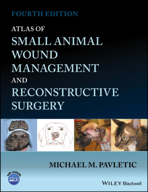
Atlas of small animal wound management and reconstructive surgery
A one-stop reference for the surgical treatment of wounds in small animal patients Wound management and reconstructive surgery are among the most challenging and innovative subspecialties of veterinary surgery for the management of traumatic injuries and neoplastic conditions commonly encountered in small animals.
eBook
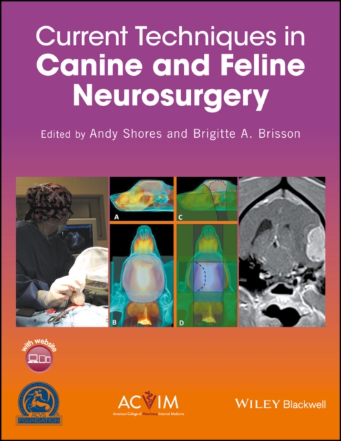
Current techniques in canine and feline neurosurgery
Current Techniques in Canine and Feline Neurosurgery offers state-of-the-art, detailed guidance on performing neurosurgical techniques in dogs and cats, from indications and surgical anatomy to procedures and post-operative care.
* Presents an up-to-date, detailed reference on veterinary neurosurgery techniques, covering skills ranging from basic to advanced
* Provides guidance on why, when, and how to perform neurosurgical procedures
* Includes information on diagnostic evaluation, surgical planning, and instrumentation as well as step-by-step descriptions of specific procedures
* Copublished with the American College of Veterinary Surgeons Foundation and American College of Veterinary Internal Medicine
* Offers video clips on a companion website
eBook
A laparoscopic approach for removal of ovarian remnant tissue in 32 dogs
Background
Surgical treatment of ovarian remnant syndrome (ORS) in dogs usually necessitates large celiotomies and considerable manipulation of organs because of the relatively deep position of ovarian remnant tissue, large patient size, and often encountered adhesions. In women, laparoscopic treatment of ORS is successful and has significant advantages over laparotomy. Since laparoscopic ovariectomy has significant advantages over open ovariectomy in dogs, including reduced surgical stress and postoperative pain and shorter convalescence period, the rationale for a laparoscopic approach of canine ORS is evident. Feasibility and efficacy of a laparoscopic approach for treatment of ORS in dogs was prospectively evaluated using a standardized protocol for diagnosis, treatment, and follow-up. Treatment success was evaluated by histology of removed tissues, postoperative hormone testing, and long-term clinical follow-up.
Results
Thirty-two client-owned predominantly medium and large breed dogs diagnosed with ORS underwent abdominal ultrasound for ovarian remnant localization prior to laparoscopic surgery for removal of ovarian remnants. Tissue dissection and excision was performed using a vessel sealing forceps. Laparoscopy subjectively enabled detailed visibility and facilitated detection and removal of suspected ovarian tissue in all cases. Histology confirmed ovarian origin of removed tissue in all dogs. Additionally, a GnRH stimulation test was performed in fourteen dogs after a median follow-up of 10.5 months, which verified absence of residual functional ovarian remnant tissue in all dogs. Median surgery duration was 97.5 min and mean total convalescence duration, subjectively scored by owners, was 1.5 ± 0.7 days. No major complications occurred. Adhesions were observed in 79% of the dogs, complicated the surgical approach, and significantly affected surgery duration (85 versus 109 min;
p
= 0.03). Minor hemorrhage occurred in 12% and significantly increased surgery duration (95.5 versus 128 min;
p
= 0.02). Trendelenburg position and lateral tilting of the patient were essential for proper access to ovarian remnants. GnRH stimulation test results and/or absence of clinical signs indicative of ORS after a median follow-up period of 22.5 months confirmed treatment efficacy in all dogs.
Conclusion
Laparoscopic surgery for ORS in dogs is effective with minimal complications and short convalescence and can successfully replace the conventional, more invasive open surgical procedure.
Journal Article

A Multicenter, Retrospective Analysis of Long-Term Survival in 255 Dogs With Pheochromocytoma Treated With Alpha-Adrenoreceptor Antagonists or Surgery (2010–2021)
Abstract
Background
The survival of dogs with pheochromocytoma (PCC) treated with adrenoreceptor antagonists has not been described or compared to surgically managed cases.
Hypothesis/Objectives
The objective of this study is to evaluate the survival of medically and surgically managed dogs with PCC and investigate factors associated with survival.
Animals
Two hundred fifty-five dogs with PCC, treated with alpha-adrenoreceptor antagonists (AA) without adrenalectomy (Group 1, n = 75), adrenalectomy +/– AA (Group 2, n = 128), or neither treatment (Group 3, n = 52).
Methods
Retrospective, multicenter review of medical records. Median overall survival time (OST) for Groups 1 and 2 combined was calculated using Kaplan–Meier estimates, and then compared between Group 1 and Group 2 using Log-Rank testing. Cox proportional hazard analysis identified factors associated with survival in Groups 1 and 2 individually and combined.
Results
Median OST for all cases was 854 (95% CI: 572–1136) days. Median OST was lower in Group 1 (247 days, 95% CI: 76–418 days) than in Group 2 (927 days, 95% CI: 587–1267 days; p < 0.001). In Group 2, 88/92 dogs (97.8%) that received presurgical AA treatment survived to discharge compared to 23/27 (85.2%) that did not receive AA pretreatment (p = 0.03). Lack of clinical signs at presentation was associated with increased survival in both groups combined (HR 0.5; 95% CI 0.3–0.9; p = 0.02) and in Group 2 alone (HR 0.3; 95% CI 0.1–0.7; p = 0.01).
Conclusions and Clinical Importance
Dogs with PCC treated with adrenalectomy have longer survival compared to those managed with AA without adrenalectomy.
Journal Article
Genome-wide based model predicting recovery from portosystemic shunting after liver shunt attenuation in dogs
Abstract
Background
In dogs with congenital portosystemic shunt (CPSS), recovery after surgical CPSS attenuation is difficult to predict.
Objectives
Our aim was to build a model with plasma albumin concentration and mRNA expression levels of hepatic gene products as predictors of recovery from portosystemic shunting after surgery.
Animals
Seventy-three client-owned dogs referred for surgical attenuation of CPSS.
Methods
A prediction model was constructed using 2 case-control studies of recovered and nonrecovered dogs after surgical CPSS attenuation. In the 1st study, a dog-specific gene expression microarray analysis was used to compare mRNA expression in intraoperatively collected liver tissue between 23 recovered and 23 nonrecovered dogs. In the 2nd study, preoperative plasma albumin concentration and the expression of microarray-selected genes were confirmed by RT-qPCR in intraoperatively collected liver samples of 31 recovered and 31 nonrecovered dogs, including 35 dogs from the 1st study.
Results
In the 1st study, 43 genes were differently expressed in recovered and nonrecovered dogs. The mean preoperative plasma albumin concentration in recovered dogs was higher compared to nonrecovered dogs (23 and 19 g/L, respectively; P = .004). The best fitting prediction model in the 2nd study included preoperative plasma albumin concentration and intraoperative DHDH, ERLEC1, and LYSMD2 gene expression levels.
Conclusion and Clinical Importance
A preclinical model was constructed using preoperative plasma albumin concentration and intraoperative hepatic mRNA expression of 3 genes that were unbiasedly selected from the genome to predict recovery from portosystemic shunting after shunt ligation. Further development is essential for clinical application.
Journal Article